Figure 2. Reduced cell survival in Li+-treated
explants. A-D: Nuclear (A, B) and
TUNEL staining (C, D) in E14.5 retinal explants
cultured in control (A, C) and Li+ (B,
D) containing medium for one day. Cell death (indicated
by TUNEL+ cells) is increased in the ganglion cell layer (arrow
in D) and in the neuroblast layer (arrowhead in D).
RGC, retinal ganglion cell layer; RPE, retinal pigment
epithelium; NB, neuroblast layer.
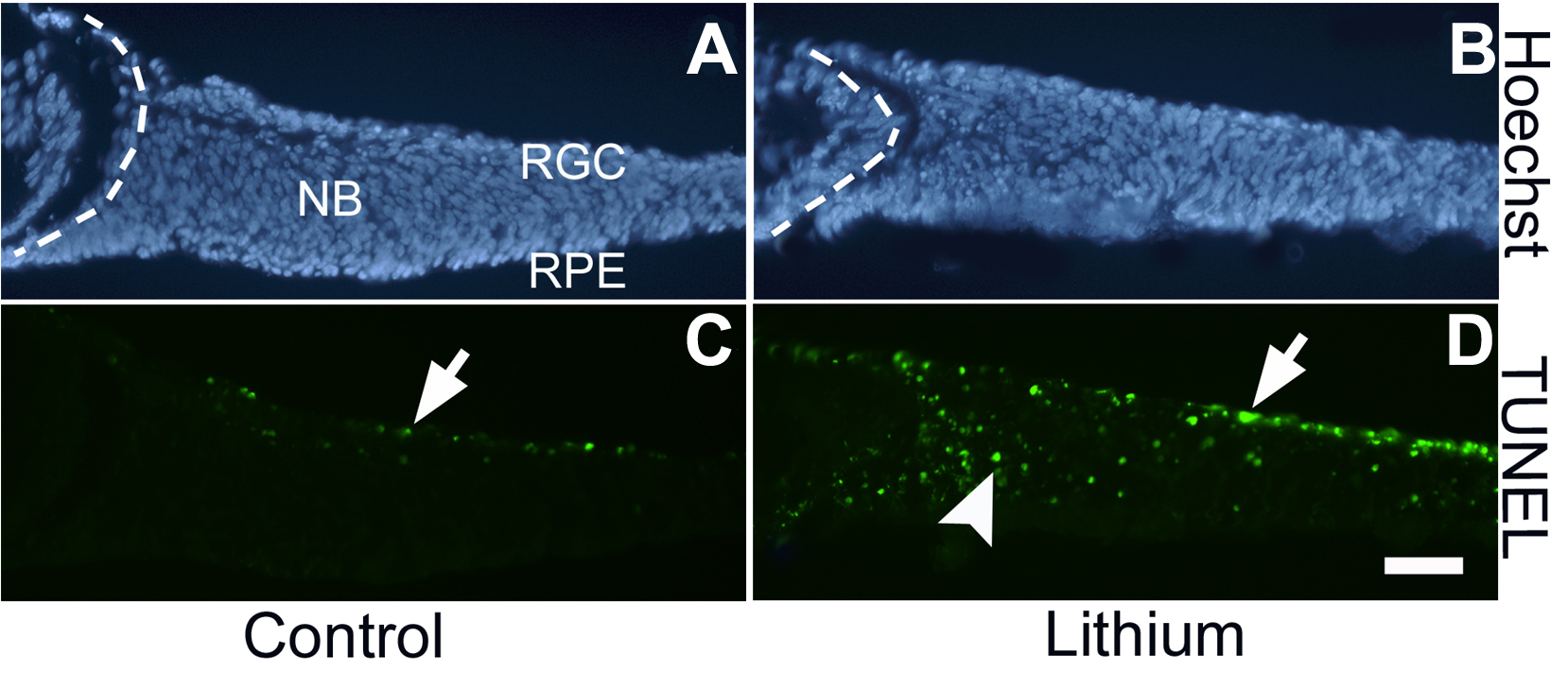
 Figure 2
of Ha, Mol Vis 2012; 18:645-656.
Figure 2
of Ha, Mol Vis 2012; 18:645-656.