Figure 7. Histopathology of corneal
grafts. Examples of grafts stained with hematoxylin-eosin on
postoperative day 14 (A-E, Magnification, 20×). A:
Untreated corneal graft (control). B: Topical
application of 1% CsA. C-E: Topical application
of 0.1% 0.3% or 0.5% FTY720, respectively. Examples of grafts
showing CD4 positive staining (brown) on postoperative day 14 (F-J,
Magnification, 40×). F: Untreated corneal graft
(control). G: Topical application 1% CsA. H-J:
Topical application of 0.1% 0.3% or 0.5% FTY720, respectively.
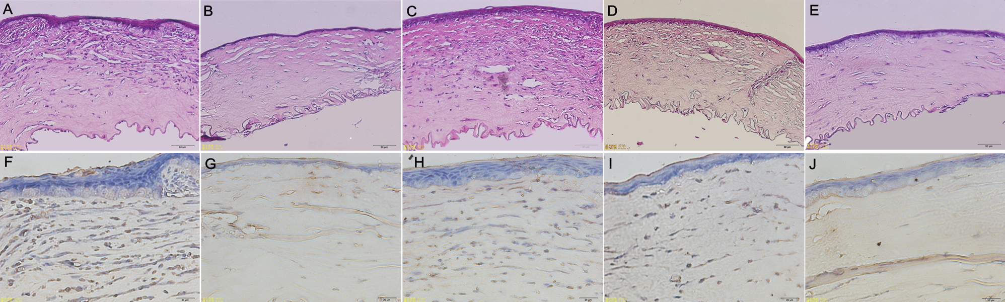
 Figure 7
of Liu, Mol Vis 2012; 18:624-633.
Figure 7
of Liu, Mol Vis 2012; 18:624-633.