Figure 2. Representative photographs
of arthritis and uveitis of patients in the family. A:
General appearance showed deformities of the feet in patient
(III:6). B: The coronal reconstruction section of CT
scan of patient (III:6) revealed erosion of bone at the edges of
the joint in the MCP joints, joint space narrowing and
subluxation in the proximal interphalangeal (PIP) joints (yellow
arrow). C: The right eye of the patient (III:6) revealed
partial posterior synechia (blue arrow). D: Deformity of
the hands of the proband (III:5). E and F:
X-rays of patient (II:5) showed multiple and symmetric joint
involvement, generalized osteoporosis, joint space narrowing,
poorly defined edges of the articular surfer, subluxation,
contracture and ankylosis of the PIP joints (white arrow). G:
the right eye of the proband (III:5) showed dot-like calcific
keratopathy in Bowman's membrane and the cornea between the
opacities remained clear (red arrow).
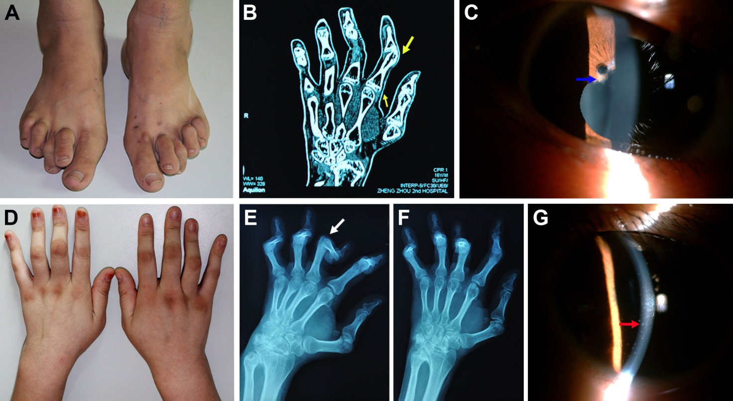
 Figure 2
of Xiang, Mol Vis 2012; 18:617-623.
Figure 2
of Xiang, Mol Vis 2012; 18:617-623.