Figure 5. Recruitment of inflammatory
cells to the cornea of Dstn mutants. A:
Immunofluorescence using CD45 (red) to mark inflammatory cells
and myeloperoxidase to mark neutrophils specifically in Dstn
mutant and WT cornea on A.BY and B6 backgrounds at P58. All
slides were counterstained with DAPI to mark cell nuclei (blue).
Bar, 20 μm. B: Quantification of CD45 positive cells
revealed significantly increased inflammation in both Dstncorn1
and Dstncorn1–2J mutant cornea compared to WT
at P58 (p<0.001 for both backgrounds). C: The trend
for increased inflammation in A.BY Dstncorn1
compared to B6.Cg-Dstncorn1 is present by P28
and increases to significance by P58. Sample sizes: A.BY.Cg-Dstncorn1-2J
P58 n=4, A.BY Dstncorn1 P58 n=8, A.BY WT P58
n=4, B6 Dstncorn1–2J P58 n=4, B6.Cg-Dstncorn1
P58 n=6, B6 WT P58 n=4, A.BY Dstncorn1 P28
n=6, B6.Cg-Dstncorn1 P28 n=3. Unit length=300
um. Error bars, SEM * denotes statistical significance resulting
from t-tests, with omitted bars representing
non-significance. *p<0.05, **p<0.01, ***p<0.001.
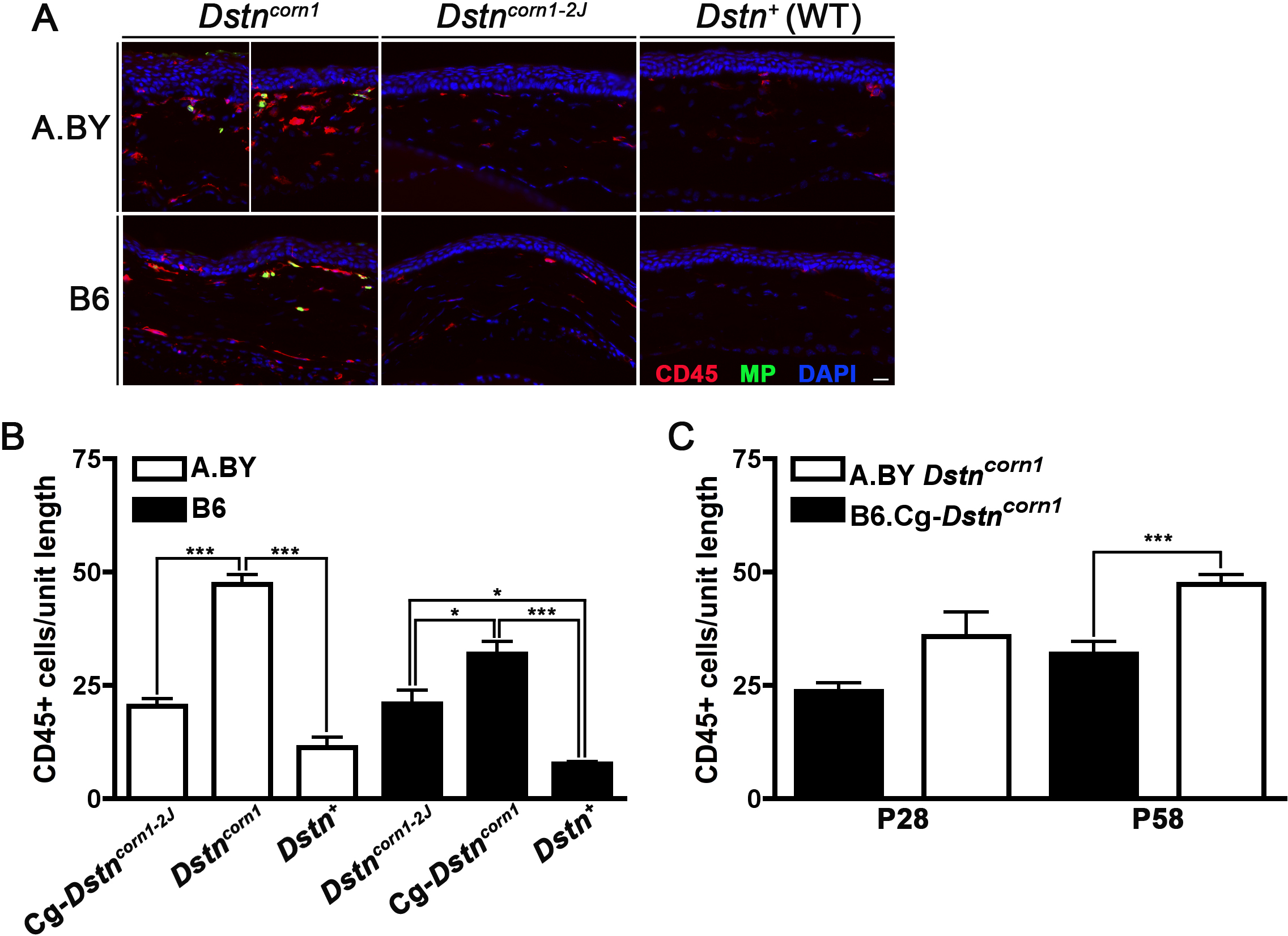
 Figure 5
of Kawakami-Schulz, Mol Vis 2012;
18:606-616.
Figure 5
of Kawakami-Schulz, Mol Vis 2012;
18:606-616.