Figure 4. Epithelial
hyperproliferation in Dstncorn1 corneas. A:
Immunofluorescence for Ki67 (green) labels proliferating cells
in the corneal epithelium of Dstn mutants and WT mice on
A.BY and B6 backgrounds at P58. All slides were counterstained
with DAPI to mark cell nuclei (blue), which is shown merged with
Ki67 staining in the lower panels. Bar, 20 μm. B:
Quantification of Ki67 positive cells shows significantly
increased numbers of proliferating cells in Dstncorn1
cornea compared to Dstncorn1–2J and WT cornea
in both A.BY and B6 backgrounds at P58 (p<0.001 for both
backgrounds). C: A tendency for increased
hyperproliferation in A.BY Dstncorn1 compared
to B6.Cg-Dstncorn1 is observed by P28. This
difference becomes statistically significant by P58. Sample
sizes: A.BY.Cg-Dstncorn1–2J P58 n=4, A.BY Dstncorn1
P58 n=5, A.BY WT P58 n=4, B6 Dstncorn1–2J P58
n=3, B6.Cg-Dstncorn1 P58 n=5, B6 WT P58 n=4,
A.BY Dstncorn1 P28 n=6, B6.Cg-Dstncorn1
P28 n=3. Unit length=300 μm. Error bars, SEM * denotes
statistical significance resulting from t-tests, with
omitted bars representing non-significance. *p<0.05,
**p<0.01, ***p<0.001.
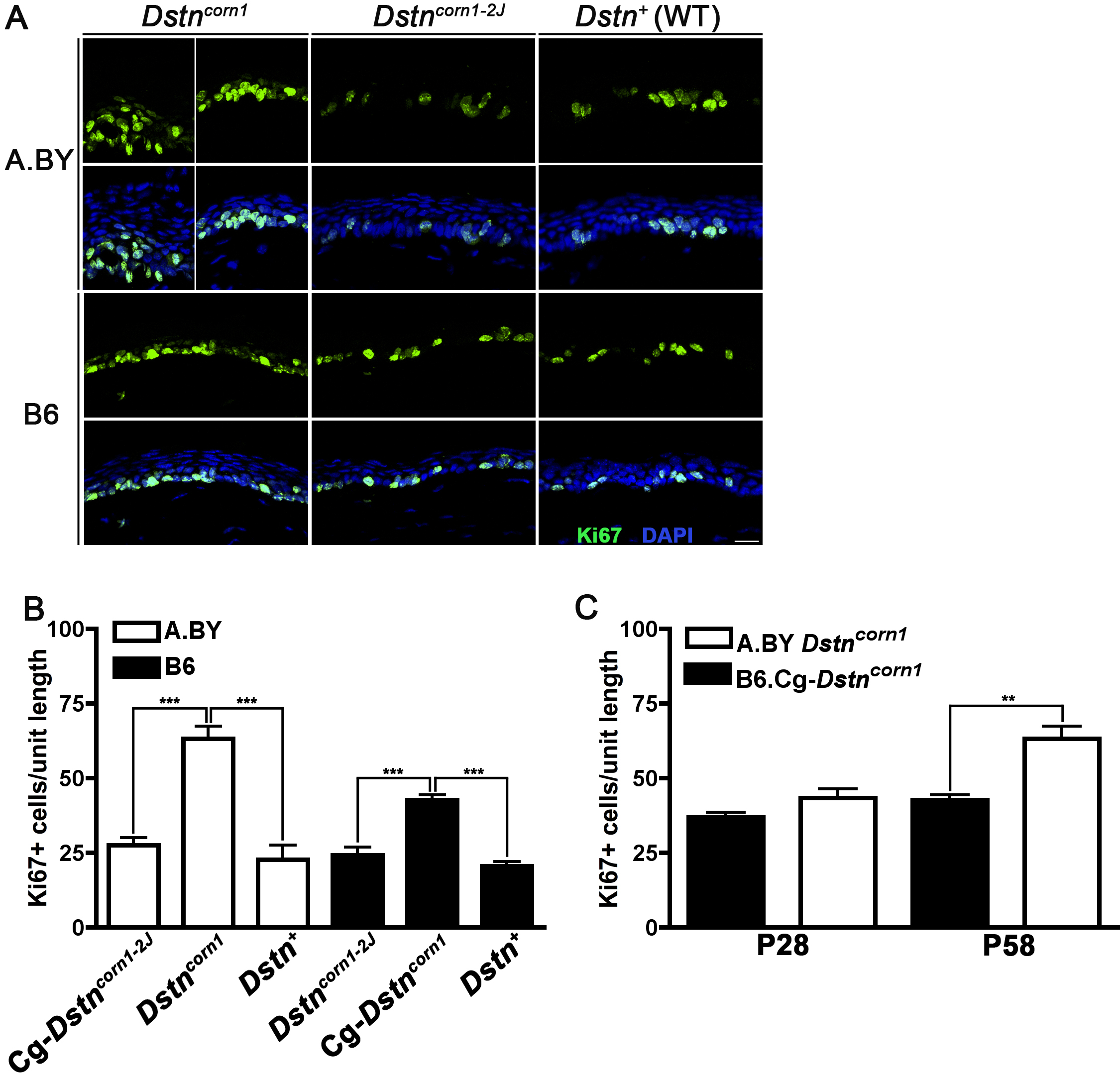
 Figure 4
of Kawakami-Schulz, Mol Vis 2012;
18:606-616.
Figure 4
of Kawakami-Schulz, Mol Vis 2012;
18:606-616.