Figure 3. Corneal neovascularization
caused by the Dstncorn1 mutation. A:
Immunofluorescence for CD31 highlights blood vessels that
infiltrate Dstncorn1 cornea in both the A.BY
and B6 background. Significant neovascularization is not
observed as a result of the Dstncorn1–2J
mutation, making the appearance similar to WT. Bar, 200 μm. B:
Quantification of the vascularized area shows that Dstncorn1
cornea have significantly more vasculature compared to Dstncorn1–2J
and WT cornea in both genetic backgrounds (p<0.001 for both
backgrounds). Note that the resting level of vasculature is
higher in B6 than A.BY. C: Genetic background effect on
the neovascularization phenotype in the Dstncorn1
cornea is significant at postnatal day 28, and becomes even more
significant with age. Sample sizes: A.BY.Cg-Dstncorn1–2J
P58 n=5, A.BY Dstncorn1 P58 n=11, A.BY WT P58
n=3, B6 Dstncorn1–2J P58 n=4, B6.Cg-Dstncorn1
P58 n=10, B6 WT P58 n=4, A.BY Dstncorn1 P28
n=11, B6.Cg-Dstncorn1 P28 n=6. Error bars, SEM
* denotes statistical significance resulting from t-tests,
with omitted bars representing nonsignficance. *p<0.05,
**p<0.01, ***p<0.001.
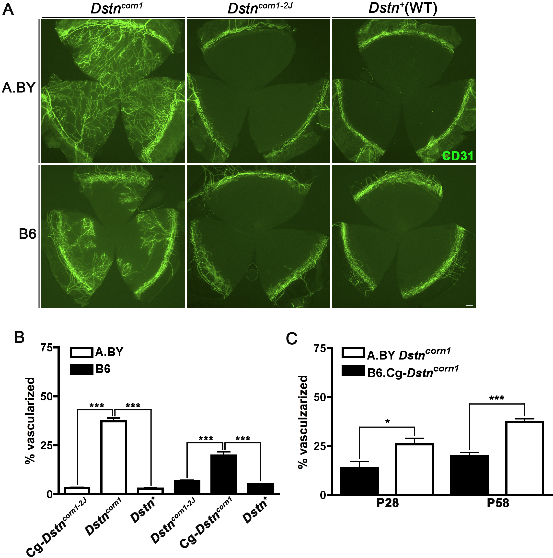
 Figure 3
of Kawakami-Schulz, Mol Vis 2012;
18:606-616.
Figure 3
of Kawakami-Schulz, Mol Vis 2012;
18:606-616.