Figure 1. Localization and expression of VAP-1 in the choroid and CNV. A and B: Representative micrographs of a laser-induced CNV lesion. (A) Phase contrast image. (B) Fluorescent micrograph of VAP-1 (red) and cell nuclei (blue). Arrowheads and arrows indicate the localization of VAP-1 in
the CNV and choroidal vessels, respectively. C: Immunoblotting analysis of VAP-1 and α-tubulin expression in the RPE-choroidal complex after laser injury.
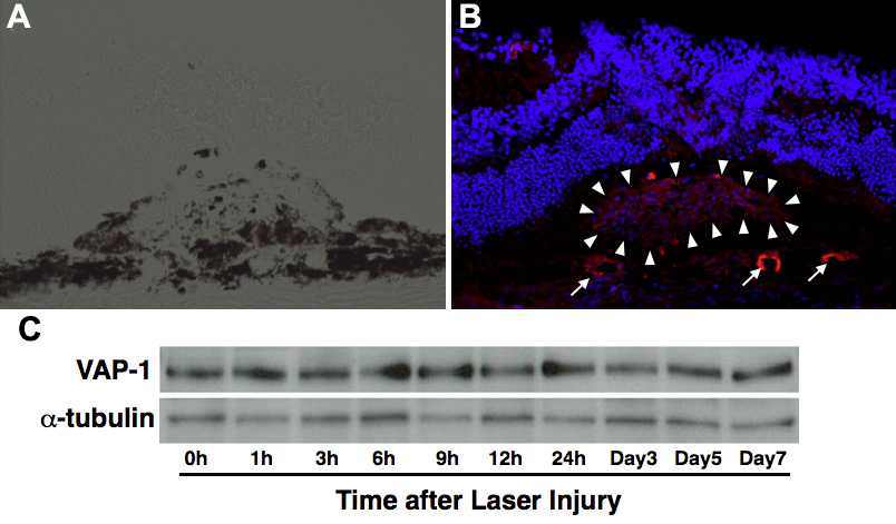
 Figure 1 of
Yoshikawa, Mol Vis 2012; 18:593-600.
Figure 1 of
Yoshikawa, Mol Vis 2012; 18:593-600.