Figure 2. CD271 stem cell marker
expression in C918 uveal melanoma cultures. The upper row of
pictures demonstrates the morphology of two-dimensional (2D; A
and C) and three-dimensional (3D; E and G)
cultures of C918 uveal melanoma with the formation of prominent
VM in 3D cultures. Arrows in E and G point to
VM. Asterisks in E and G point to cells growing
in monolayer on the Matrigel surface. Arrowheads in E
and G point to cells growing on the bottom of the
culture dish. Panel H demonstrates detection of CD271
expression by VM-forming tumor cells by immunofluorescence in 3D
cultures. No CD271 expression was detected in 2D cultures (D).
CD271 expression was also not detected in control 2D and 3D
cultures where no primary antibody was used (B and F).
Magnification: 200×.
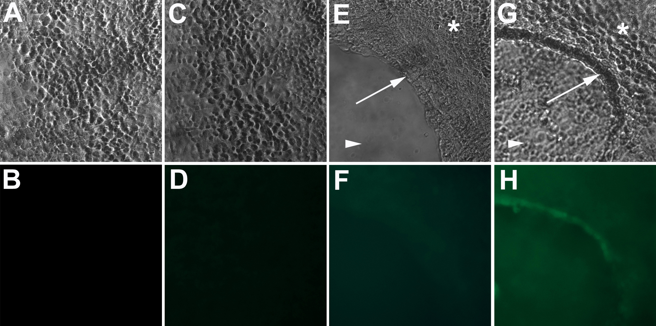
 Figure 2
of Valyi-Nagy, Mol Vis 2012; 18:588-592.
Figure 2
of Valyi-Nagy, Mol Vis 2012; 18:588-592.