Figure 1. Morphology of 3D C918 uveal
melanoma cultures. Pictures of a 3D C918 uveal melanoma culture
with focus either on cells growing on the Matrigel surface (A)
or on cells growing on the bottom of the tissue culture well (B).
Vasculogenic mimicry (VM) patterns are marked by arrows,
asterisks point to cells growing in monolayer on the Matrigel
surface and arrowheads highlight cells growing on the bottom of
the culture dish. Magnification: 200×.
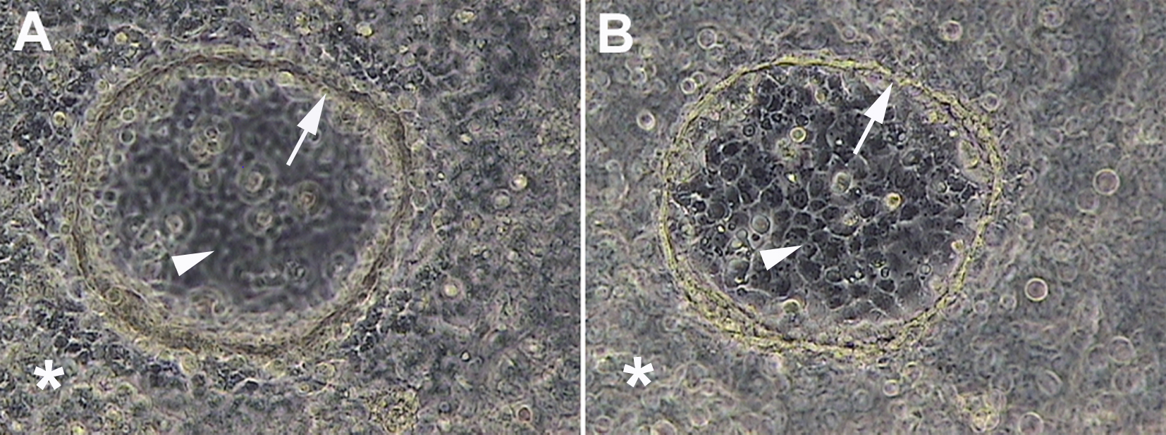
 Figure 1
of Valyi-Nagy, Mol Vis 2012; 18:588-592.
Figure 1
of Valyi-Nagy, Mol Vis 2012; 18:588-592.