Figure 4. Trafficking studies of the mutant protein. A: Subcellular localization of the WT opsin-GFP, P23H opsin-GFP and the novel c.Y206-F208del opsin-GFP mutation in SK-N-SH
neuroblastoma cells. The arrows indicate plasma membrane localization for the WT opsin and inclusion bodies for both mutant
proteins. The scale bar represents 10 μm. B: Quantification of the predominant subcellular localization in SK-N-SH neuroblastoma of the WT opsin-GFP, P23H opsin-GFP,
and Y206-F208del opsin-GFP fusion proteins 24 h after transfection. Three batches of 100 cells were counted and predominant
localization of the opsin in the plasma membrane (PM), endoplasmic reticulum (ER), or the presence of an inclusion were graded.
Error bars represent ±2 standard errors.
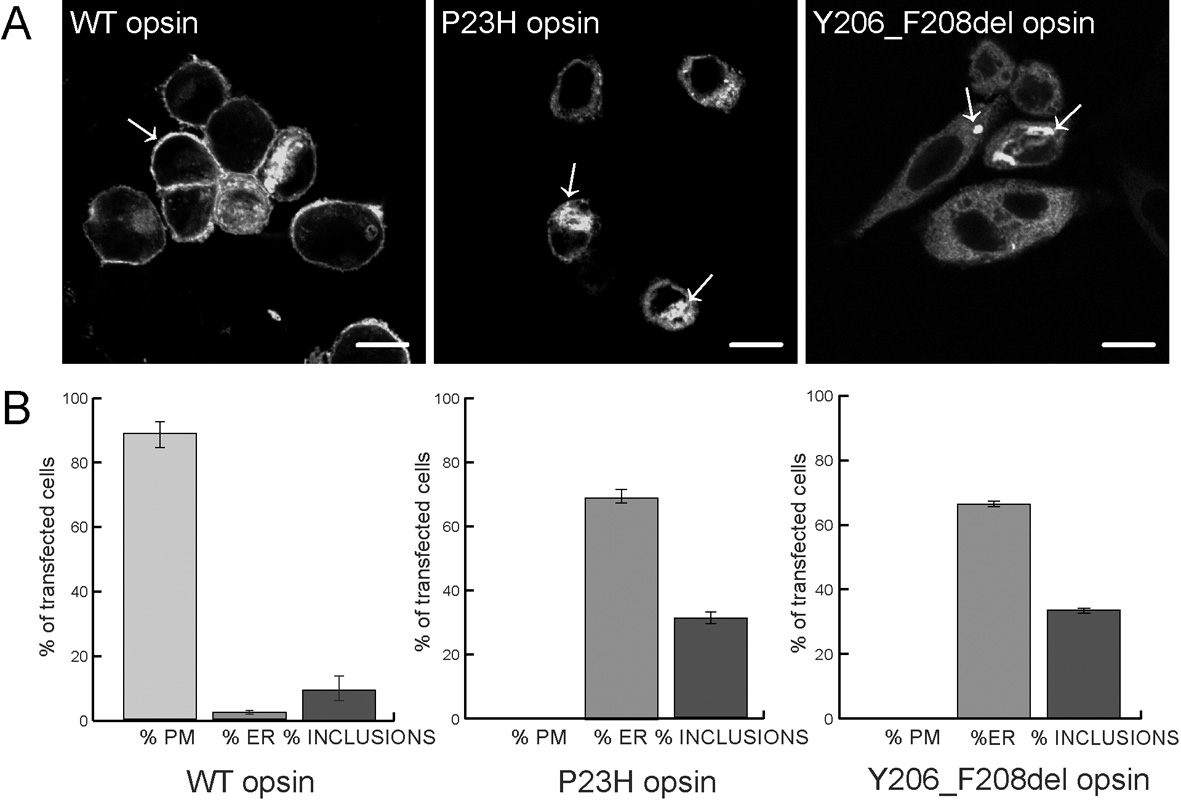
 Figure 4 of
Maubaret, Mol Vis 2012; 18:581-587.
Figure 4 of
Maubaret, Mol Vis 2012; 18:581-587.