Figure 2. Clinical information on the proband. A: Fundus photography of the right eye shows classical bony-spicules, attenuated vessels, and waxy pale optic discs. B: Results of a120-point visual field screening showing severe constriction of both visual fields. In this automatic visual
field examination (screening test), visual stimuli were presented in different points of the visual field (throughout a virtual
map shown here). White circles (in the center) represent stimuli detected by the proband, whereas black rectangles correspond
to undetected stimuli.
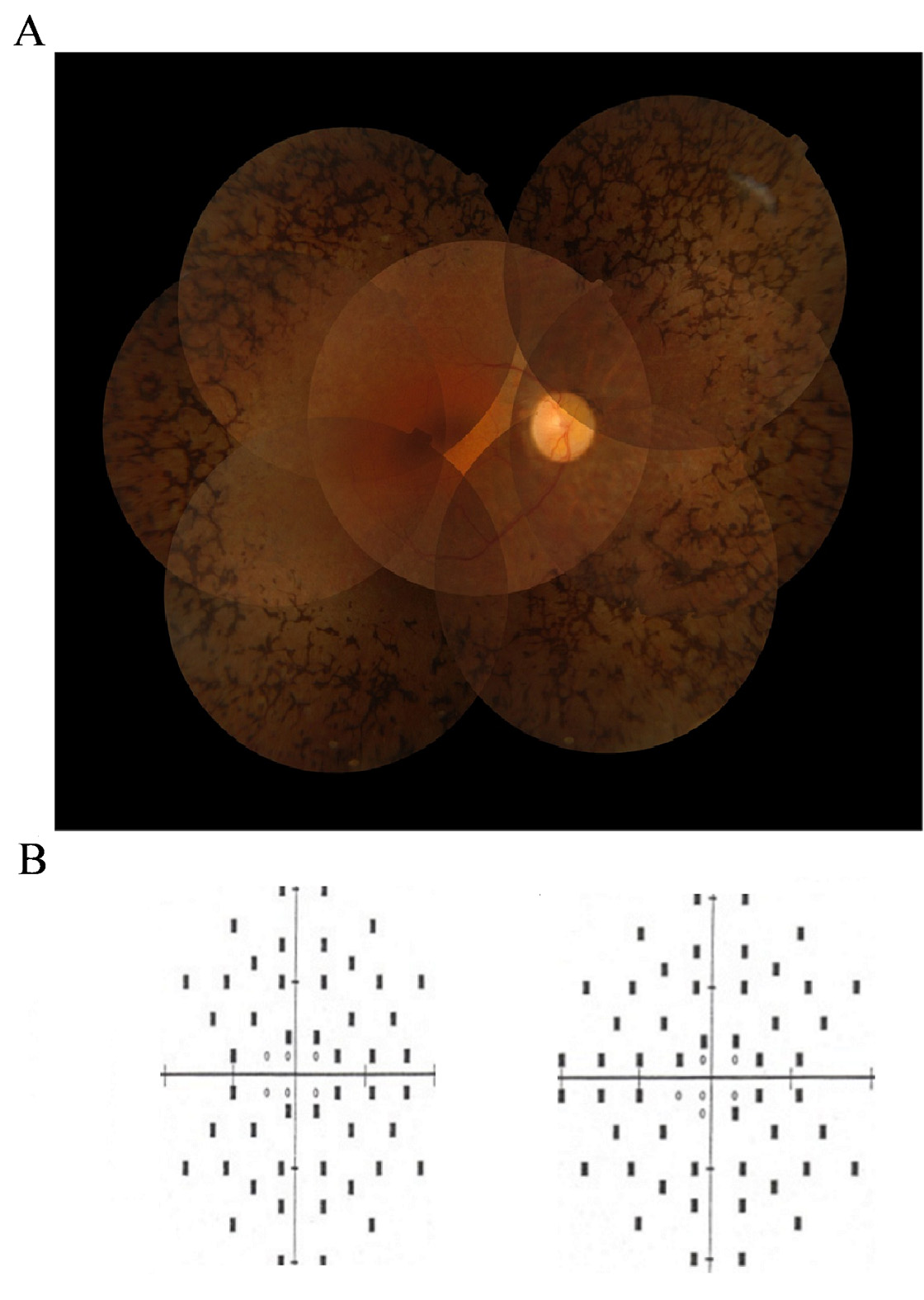
 Figure 2 of
Maubaret, Mol Vis 2012; 18:581-587.
Figure 2 of
Maubaret, Mol Vis 2012; 18:581-587.