Figure 7. Attachment of GFP-labeled
UCB1 MSCs to damaged endothelium. The image in (A) shows
lack of attachment of UCB1 MSCs to unwounded endothelium. The
inset shows results of the no-primary negative control for ZO1
staining. GFP-labeled UCB1 MSCs in (B) attached to
damaged endothelium in the crush wound model. Arrowheads
indicate the ZO1 pattern of unwounded HCEC. The image in (C)
shows attachment of GFP-labeled UCB1 MSCs to remnants of damaged
endothelium in the scrape wound model. Arrows indicate areas of
damaged HCEC. Red: ZO1. Blue: TO-PRO-3-stained nuclei. Blue:
TO-PRO-3-stained nuclei. Original magnification: 40×.
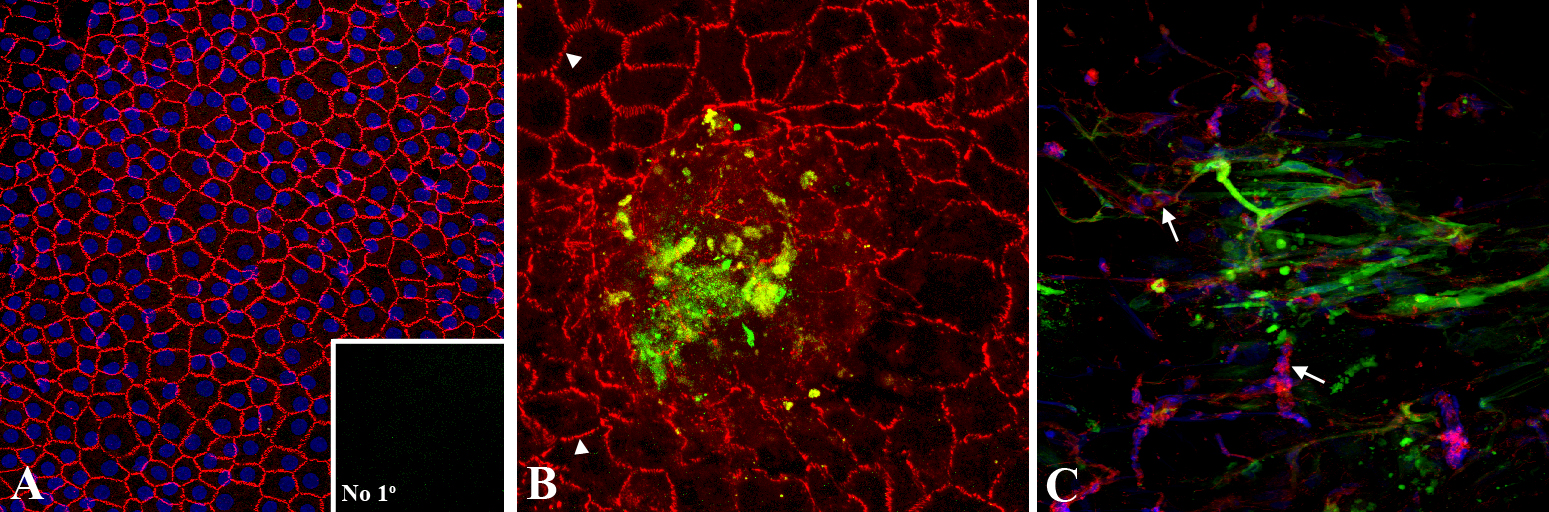
 Figure 7
of Joyce, Mol Vis 2012; 18:547-564.
Figure 7
of Joyce, Mol Vis 2012; 18:547-564.