Figure 6. Effect of EGTA treatment on UCB1 MSC morphology and junction-associated protein localization. Top four phase-contrast images
demonstrate that EGTA treatment induces separation of UCB1 MSCs in cultures grown in either lens epithelial cell basal medium
(LECBM) or lens epithelial cell-conditioned medium (LECCM). Arrows in the (+) EGTA images indicate large spaces between cells.
Confocal fluorescence images at the bottom demonstrate changes in the relative localization of N-cadherin (FITC) and ZO1 (rhodamine)
in UCB1 MSCs grown in LECCM. Both bottom images are overlays with TO-PRO-3 (blue) to visualize nuclei. Phase contrast original
magnification: 4×. Confocal original magnification: 40×.
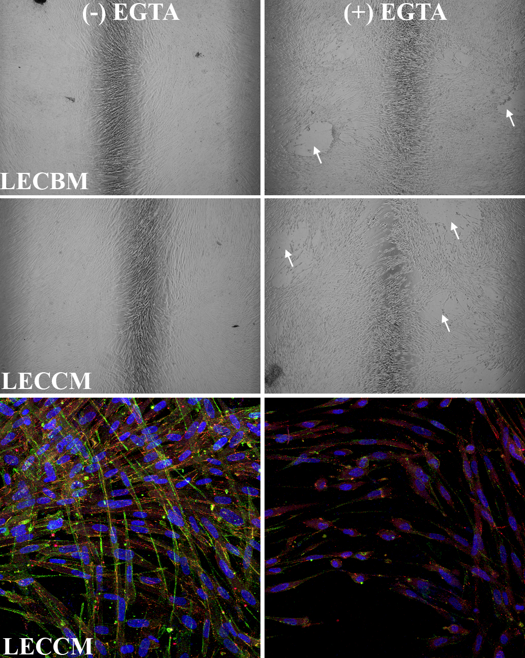
 Figure 6 of
Joyce, Mol Vis 2012; 18:547-564.
Figure 6 of
Joyce, Mol Vis 2012; 18:547-564.