Figure 1. Phase-contrast images of
UCB1 and UCB4 MSCs. Note the more elongated shape and swirl
pattern formed by UCB1 cells compared with the broader shape of
UCB4 cells growing in focal patches. UCB1 MSCs are passage 11.
UCB4 MSCs are passage 3. Original magnification of (A)
and (B): 4×. Original magnification of (C) and (D):
10×.
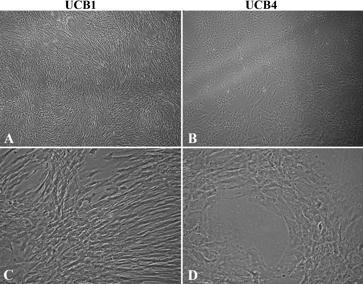
 Figure 1
of Joyce, Mol Vis 2012; 18:547-564.
Figure 1
of Joyce, Mol Vis 2012; 18:547-564.