Figure 1. Expression of TYR,
MITF-A, MITF-D, MITF-H, MITF-M,
and OTX2 mRNA in RPE cells. ARPE-19 mRNA was collected
at various time points (6 h, 24 h, 48 h, 96 h, 14 days, and 30
days). Other samples were isolated at 96 h from D407 RPE cells
and G-361 melanoma cells, a positive control for TYR
expression. Results are presented as the mean normalized
expression±SD relative to 6 h ARPE-19 culture (=1) from three to
four independent cultures each performed in triplicate. Due to
very low expression levels, MITF-D values were
normalized relative to MITF-H (6 h ARPE-19 culture=1).
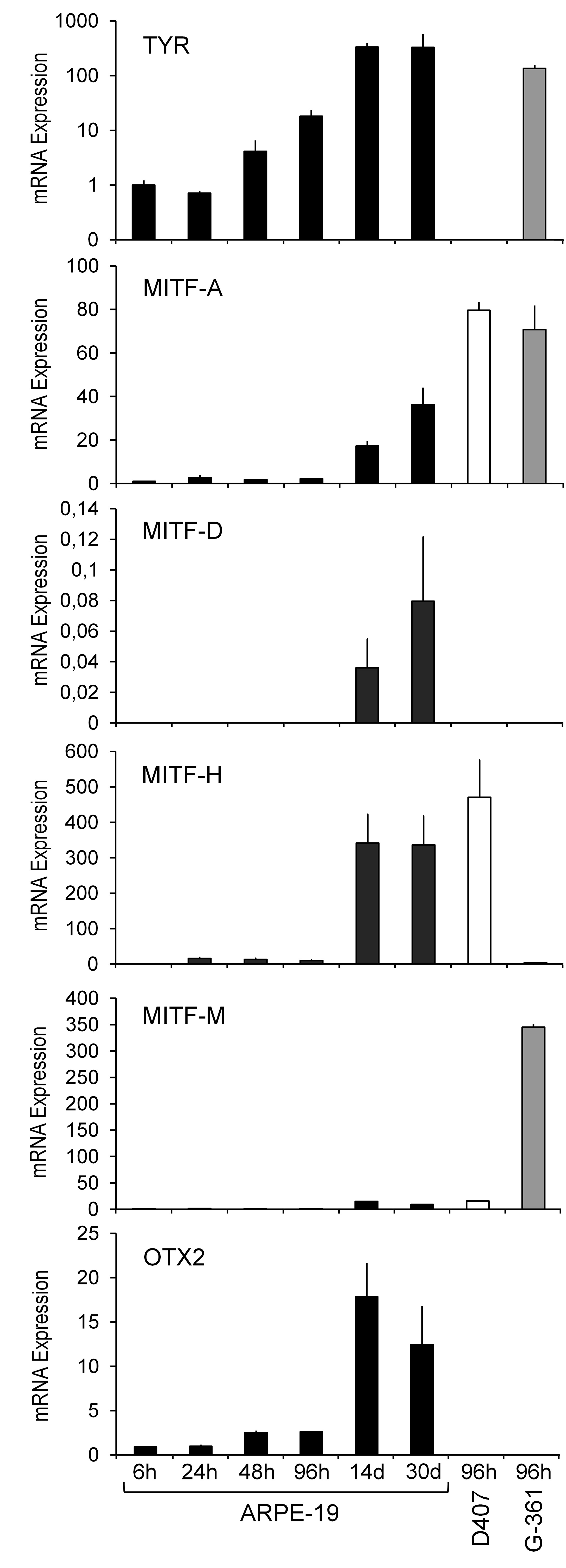
 Figure 1
of Reinisalo, Mol Vis 2012; 18:38-54.
Figure 1
of Reinisalo, Mol Vis 2012; 18:38-54.