Figure 6. c-Met was a target of
microRNA-34 b/c. A: Two specific binding sites of
miR-34b/c in the c-Met 3′ untranslated region (UTR) was
marked with black color. Alignment between the predicted
miR-34b/c target sites and miR-34b/c, the common 8 bp seed
sequence for miR-34b/c:mRNA (mRNA) pairing is shown. B:
Design of the pMIR luciferase reporter constructs, containing
c-Met 3′ UTR, which was used to verify the putative miR-34b/c
binding sites. C: SP6.5 cells were cotransfected with
miR-34b/c, pLuc-MET 3′ UTR, and a pRL-SV40 reporter plasmid. The
luciferase activity was measured after 24 h. Values are
presented as relative luciferase activity after normalization to
Renilla luciferase activity. *: Differences in luciferase
activity between miR-34b/c and negative control transfected
cells were significant, p<0.01.
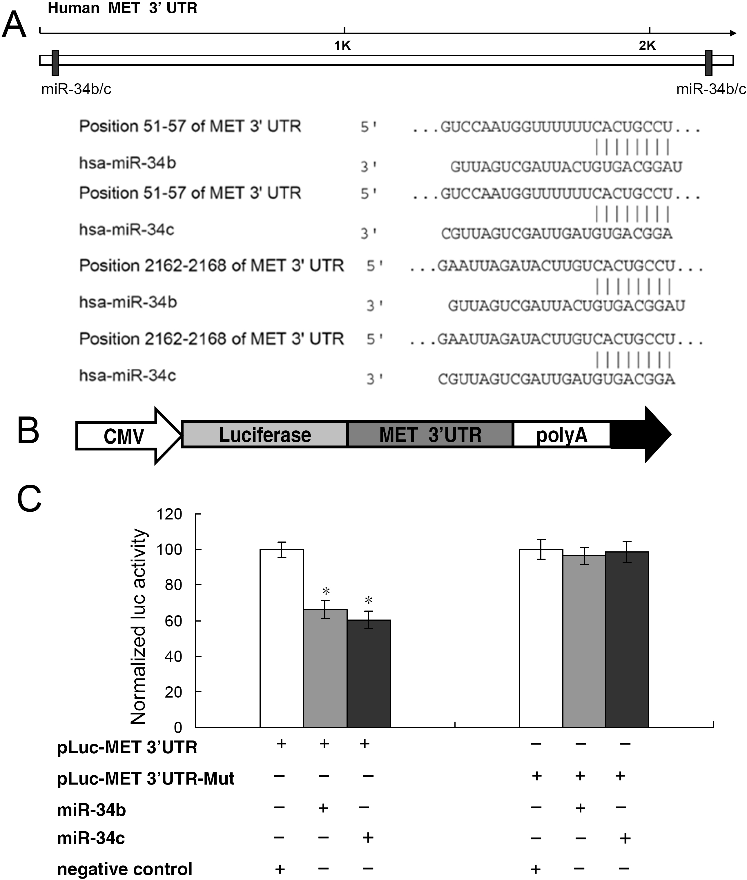
 Figure 6
of Dong, Mol Vis 2012; 18:537-546.
Figure 6
of Dong, Mol Vis 2012; 18:537-546.