Figure 3. Retinal pigment epithelium
(RPE) secretion of vascular endothelial growth factor (VEGF)
during in vitro polarization. Conditioned media were collected
from the apical and basolateral Transwell compartments over the
course of 4 weeks and analyzed for VEGF with enzyme-linked
immunosorbent assay (ELISA). A: Diagram illustrating
Transwell system used for culture of human retinal pigment
epithelium (ARPE-19) cells. B: An increase in VEGF
secretion preferentially toward the basolateral side was
observed during the 4-week culturing. The experiment was
performed in triplicate, and the results are expressed as the
mean±standard deviation.
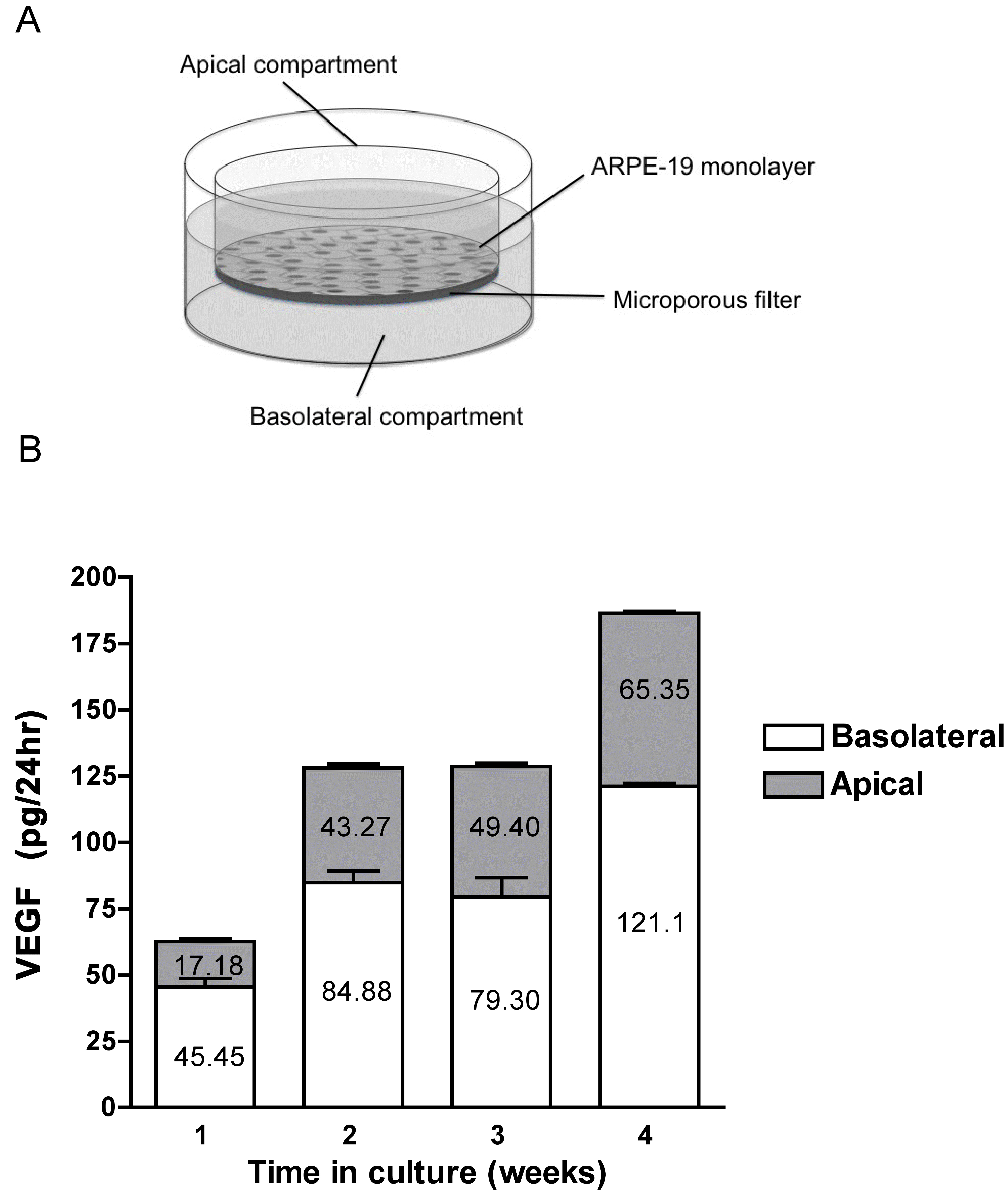
 Figure 3
of Ford, Mol Vis 2012; 18:519-527.
Figure 3
of Ford, Mol Vis 2012; 18:519-527.