Figure 4. Halofuginone treatment down-regulates Smad3 protein expression but not relative Smad3 phosphorylation in human corneal fibroblasts.
A: Representative western blots show halofuginone (Hal) reduces Smad3 protein expression. While Smad2 phosphorylation (P-Smad2)
is not affected by halofuginone, the intensity of phosphorylated Smad3 (P-Smad3) signal decreases significantly. β-actin and
GAPDH shown as simultaneous loading controls. B: Quantification and normalization of Smad2, phospho-Smad2, Smad3, and phospho-Smad3 western blot data. * p<0.05 compared
to control, ** p<0.05 compared to TGF-β (n=3, bar=S.E.M.). C: Dose-dependent down-regulation of Smad3 protein by halofuginone treatment for 24 h before addition of TGF-β. D: Time-dependent down-regulation of Smad3 protein by 10 ng/ml halofuginone treatment.
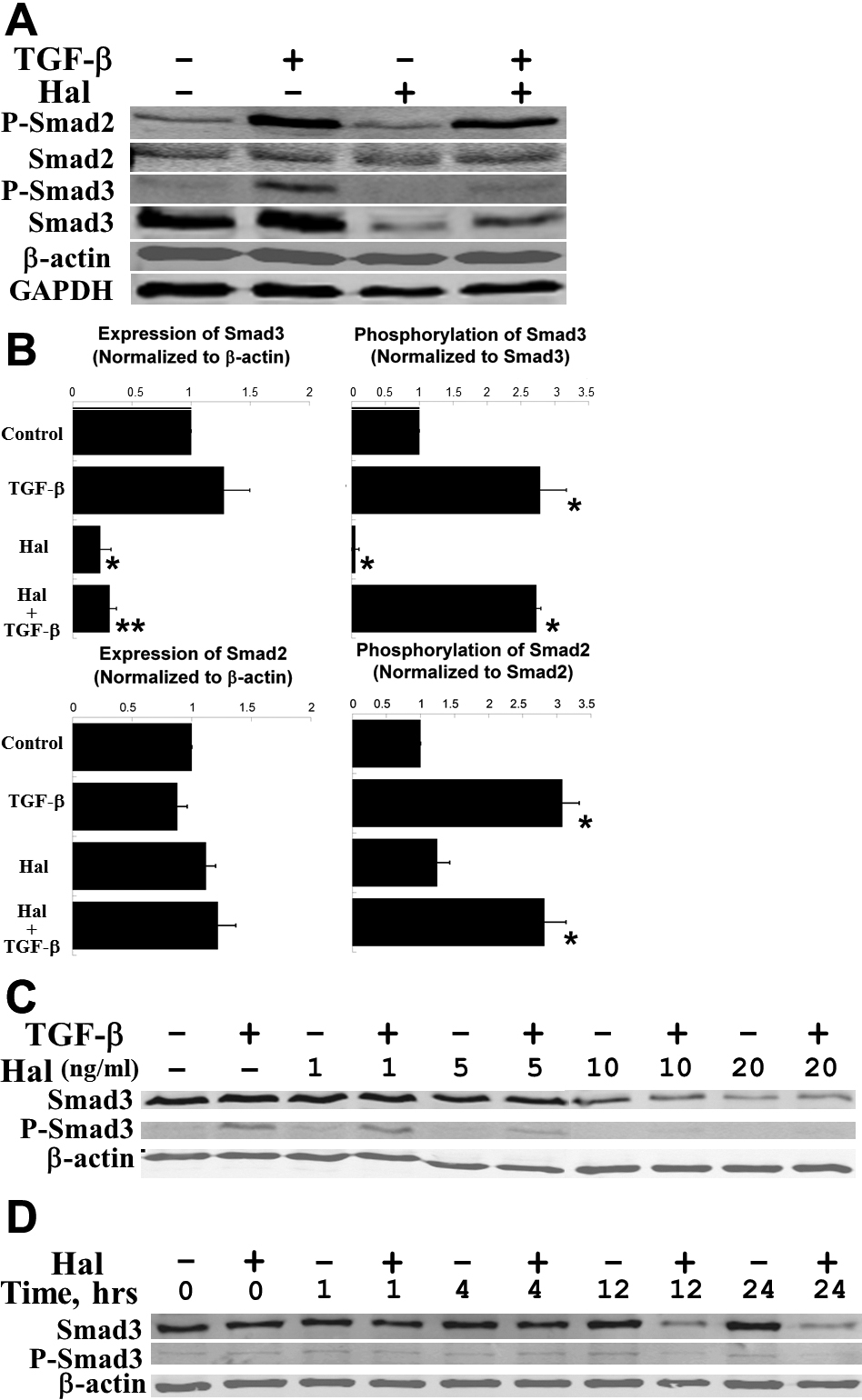
 Figure 4 of
Nelson, Mol Vis 2012; 18:479-487.
Figure 4 of
Nelson, Mol Vis 2012; 18:479-487.