Figure 2. Halofuginone suppresses TGF-β-induced expression of fibrotic markers and stress fiber assemblies in human corneal fibroblasts.
A: Representative western blots show increased expression of α-SMA and fibronectin (FN) after TGF-β treatment and inhibition
of the induced expression by halofuginone (Hal). β-actin shown as loading control. B: Quantification and normalization of α-SMA and fibronectin western blot data. * p<0.05 compared to control, ** p<0.05 compared
to TGF-β (n=4, bar=S.E.M.). C: Representative images of phalloidin toxin staining show cells treated with TGF-β (top right) display greater fluorescence
signal than untreated control (top left) or cells treated with TGF-β and halofuginone (lower right).
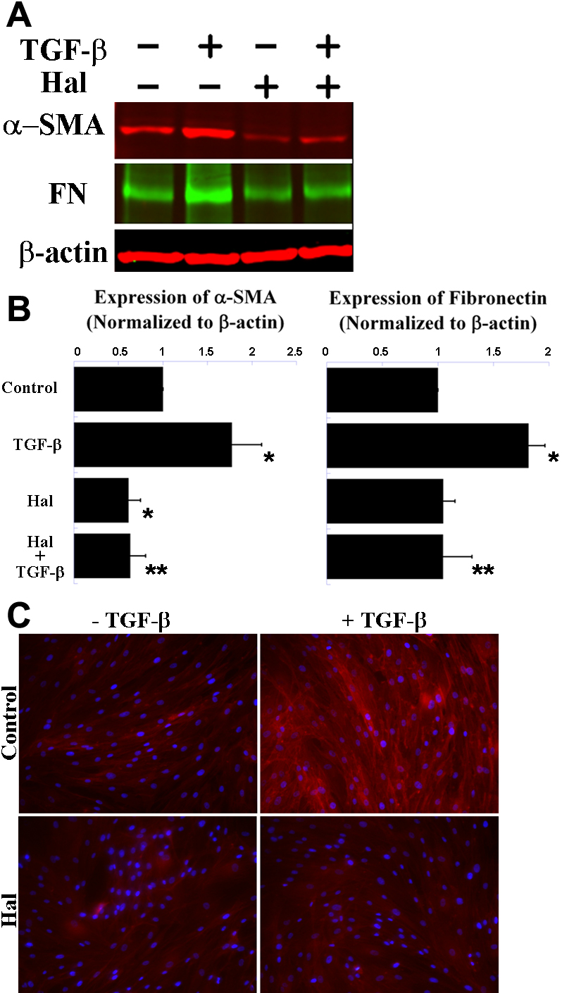
 Figure 2 of
Nelson, Mol Vis 2012; 18:479-487.
Figure 2 of
Nelson, Mol Vis 2012; 18:479-487.