Figure 4. Fundus photograph of the
right eye of Patient 15 at age 38. Note the mild pallor of the
optic disc, the attenuation of the vessels, the chorioretinal
coloboma, the retinal pigment epithelium atrophy (RPE) atrophy,
and the bone spicule-like pigmentations.
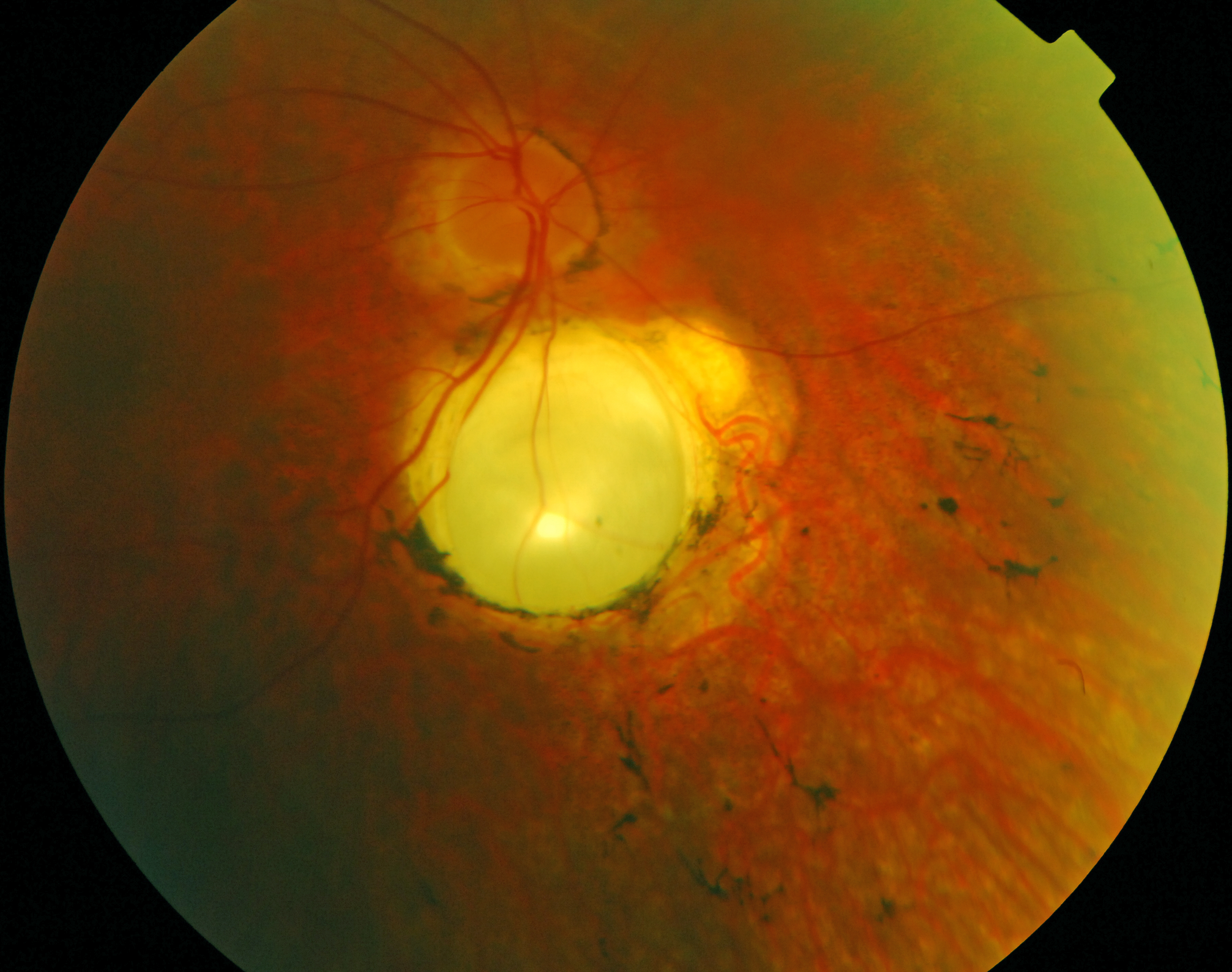
 Figure 4
of Yzer, Mol Vis 2012; 18:412-425.
Figure 4
of Yzer, Mol Vis 2012; 18:412-425.