Figure 3. Fundus photographs and OCT of Patient 7 at age 39. A: Note the pseudopapillary edema, vascular sheathing, preserved RPE in the posterior pole, subtle RPE changes in the macula,
and the pronounced RPE and choriocapillary atrophy with bone spicule-like pigmentations along the vascular arcade in the right
eye. B: Note the exudation in the posterior pole with shallow detachment in the left eye. C: Spectralis optical coherence tomography (OCT) image of the macular region of the right eye showing cyst-like changes (*)
in the outer nuclear layer with preservation of the inner/outer segment junction under the fovea but absent in the parafoveal
region. Note the thinning of the outer nuclear layer at the center (arrow).
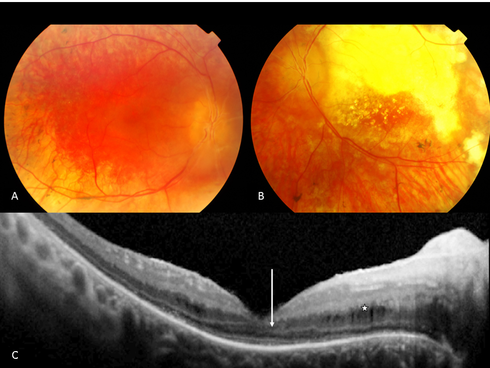
 Figure 3 of
Yzer, Mol Vis 2012; 18:412-425.
Figure 3 of
Yzer, Mol Vis 2012; 18:412-425.