Figure 2. Fundus photographs of the right eye of Patient 3 at age 13. A: Note the pink optic disc with scleral rim, mild attenuation of vessels, and preservation of the RPE in the posterior pole,
with greyish, darker appearance of the macula with subtle wrinkling of the inner limiting membrane. B: Note the subtle, atrophic, well defined dot-like changes at the level of the RPE and the bone spicules in the periphery.
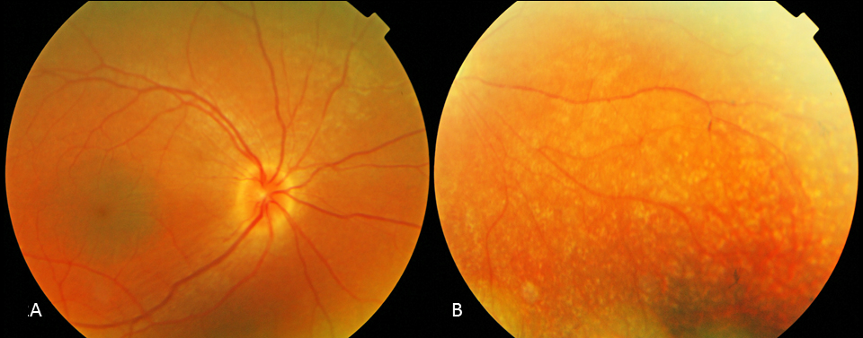
 Figure 2 of
Yzer, Mol Vis 2012; 18:412-425.
Figure 2 of
Yzer, Mol Vis 2012; 18:412-425.