Figure 4. Increased cell
proliferation at earlier responses to wounding in K14-cre+/−/NICD+
cornea. A: Quantification of BrdU positive cells in the
corneal epithelium of the WT and K14-cre+/−/NICD+
mice at different time points after wounding. The average number
of the BrdU positive cells per 1 mm length parallel to the
cornea surface was calculated from the counts on three random
sections of each four wounded eyes. B: Quantification of
BrdU positive cells in the limbal epithelium of the WT and K14-cre+/−/NICD+
mice at different time points after wounding. Each bar
represents the average number of the BrdU+ cells per
1 mm limbal epithelium±SD. The number was calculated from the
counts on three random sections from each four wounded eyes for
each experimental group. Two-tail t-test: **p<0.005
and *p<0.05. n=12.
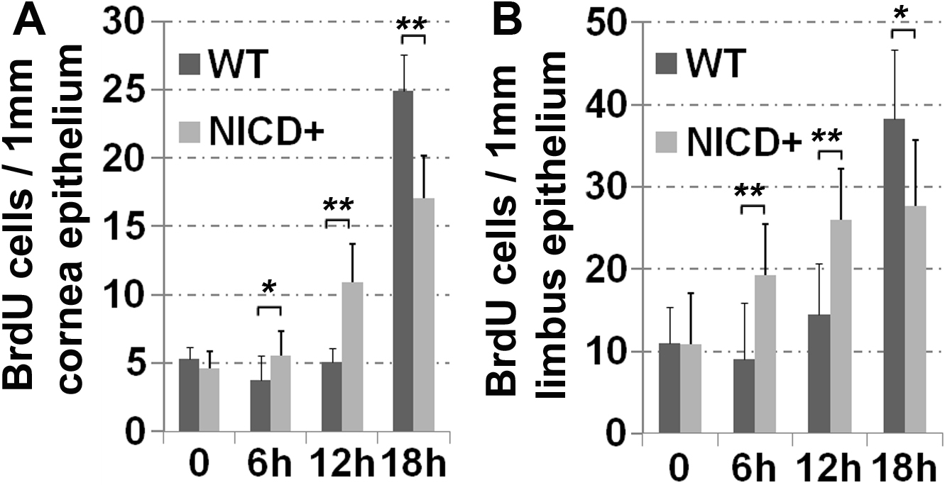
 Figure 4
of Lu, Mol Vis 2012; 18:403-411.
Figure 4
of Lu, Mol Vis 2012; 18:403-411.