Figure 3. Fast recovery from cornea
wounding in K14-cre+/−/NICD+
transgenic mice. A: The fluorescein stained eye images
were taken under cobalt-filtered ultraviolet light right after,
or 18 h after, mechanical scraping of the center corneal
epithelium. B: Quantitative measurement of wounding area
stained by fluorescein is presented as average wound area per
eye. “N” in the figure represents the number of eyes examined.
Two-tail t-test: *p<0.05. C: Wounded corneas
were stained with Richardson’s solution to show the remaining
wound area (blue at 0, 18, and 24 h after injury). D:
The wound size (Richardson stained region) was measured using
photographs with computer image software. Four eyes for each
group and the results are presented as average±SD.. Two-tail t-test:
**p<0.005 and *p<0.05. n=4.
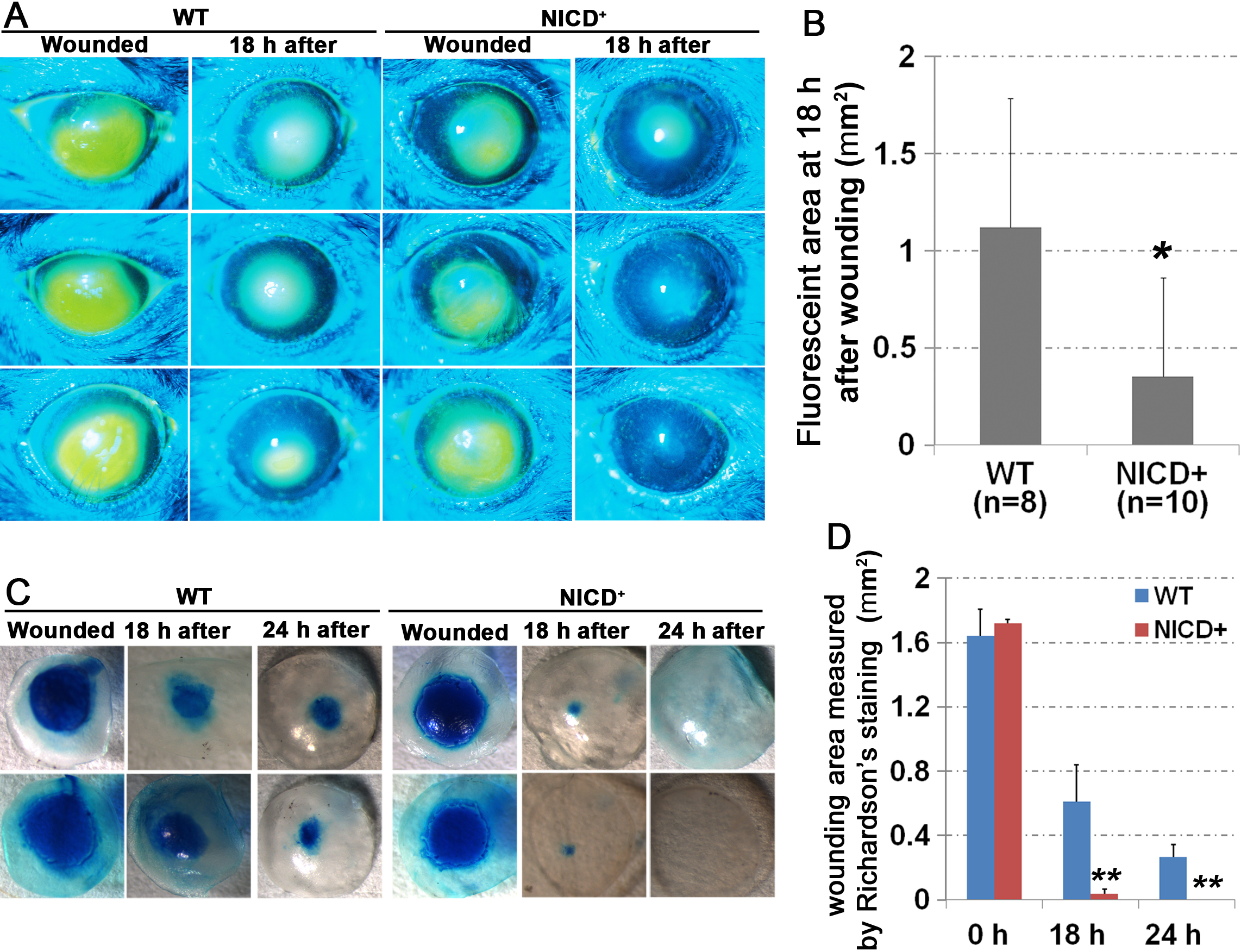
 Figure 3
of Lu, Mol Vis 2012; 18:403-411.
Figure 3
of Lu, Mol Vis 2012; 18:403-411.