Figure 2. No development defects in NICD
transgenic cornea. A: Immunohistochemistry examination
of NICD transgenic eye in comparison with wild type
control animal. The representative images of HE staining and
immunofluorescent staining with antibodies recognizing K14, p63,
BrdU, K12, and ZO-1 in the corneas of 8-weeks-old WT and K14-cre+/−/NICD+
mice. The scale bar represents 50 μm of length. B:
Quantitative examination showed no significant difference in the
cell proliferation between NICD transgenic cornea and WT
cornea. The average numbers of the BrdU positive cells per 1 mm
of fixed length parallel to the cornea surface or limbus±SD were
calculated from 5 eyes for each type of mice. n=5.
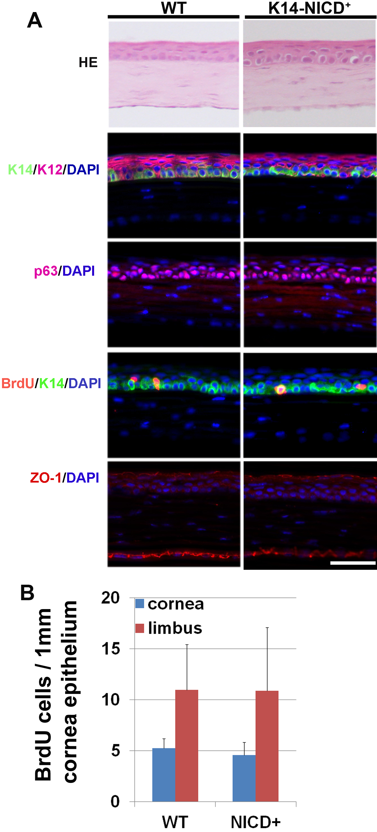
 Figure 2
of Lu, Mol Vis 2012; 18:403-411.
Figure 2
of Lu, Mol Vis 2012; 18:403-411.