Figure 2. Retinas after oxygen
induced retinopathy (OIR) protocol. A: Flatmounted
retina of a BALB/cByJ mouse on postnatal day 16 after exposure
to the OIR protocol. The retina is relatively well vascularized
at this time point. B: A retina from a C57BL/6ByJ mouse
at the same time point after exposure to the OIR protocol. The
central retina has large areas of avascularity.
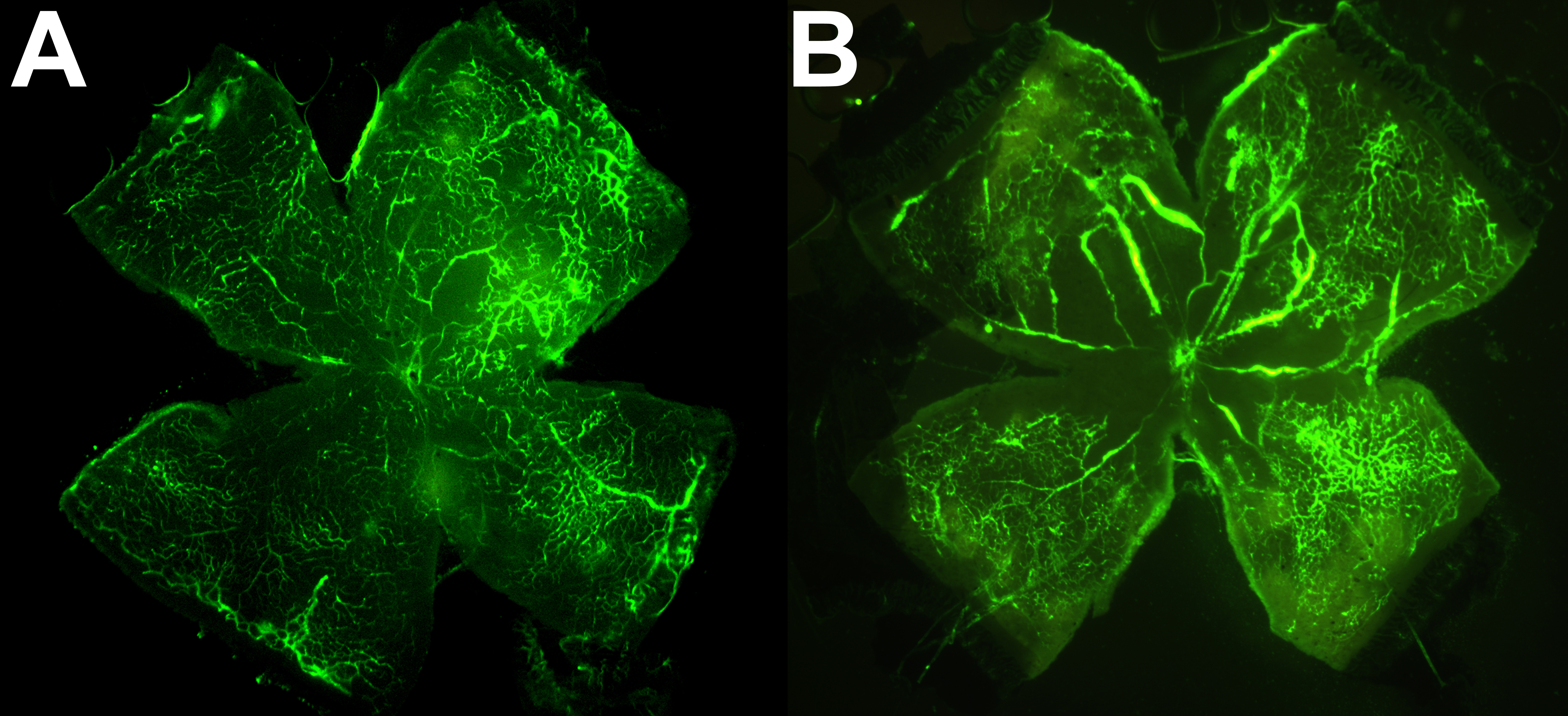
 Figure 2
of O’Bryhim, Mol Vis 2012; 18:377-389.
Figure 2
of O’Bryhim, Mol Vis 2012; 18:377-389.