Figure 5. Histopathology. A: The corneal specimen from the proband of the NTUH-18 family showed irregular thickness of the epithelium, vacuolization
in the basal epithelium, and focal subepithelial fibrosis interposed between the irregular epithelium with a “sawtooth-like”
configuration (PAS staining, 200×). B: Focal disruption of Bowman’s membrane (arrows) was replaced by subepithelial fibrotic tissue (star) (H&E staining, 400×).
C: The specimen from the proband of the NTUH-11 family showed several eosinophilic deposits interspersed within the entire
corneal stromal layer (H&E staining, 200×). D: These deposits showed green birefringence under a polarized microscope (Congo red staining, 200×).
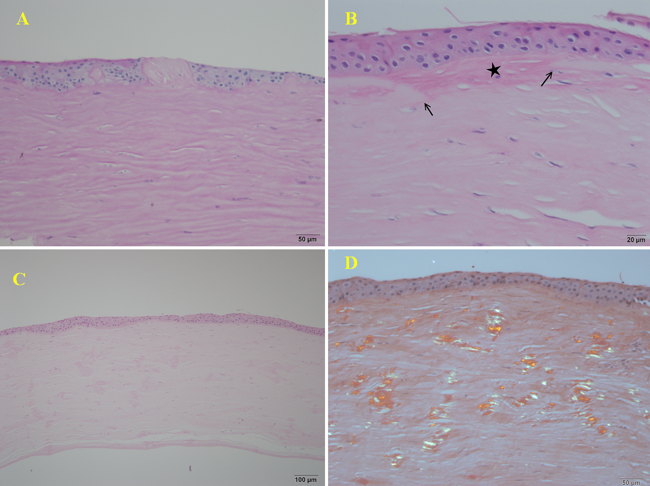
 Figure 5 of
Hou, Mol Vis 2012; 18:362-371.
Figure 5 of
Hou, Mol Vis 2012; 18:362-371.