Figure 3. Clinical phenotypes of TGFBI corneal dystrophies. A: Four round white opacities in the proband of the NTUH-2 family. B: Numerous crumb-shaped opacities in the proband of the NTUH-1 family. C: Numerous sand-like opacities with some rod-dot granules in the LASIK flap interface in the proband’s younger sister in the
NTUH-2 family. D: Some dots with thin lines in the proband of the NTUH-6 family. E: Superficial breadcrumb-like opacities in the proband of the NTUH-4 family. F: Superficial reticular opacities in the proband of the NTUH-18 family. G: Flake-dot opacities with lattice-line opacities in the proband of the NTUH-9 family. H: Superficially central diffuse haze with some very fine and short lines in the periphery in the proband of the NTUH-15 family.
I: Numerous small, polymorphic dots with some filamentous lines in the proband of the NTUH-11 family.
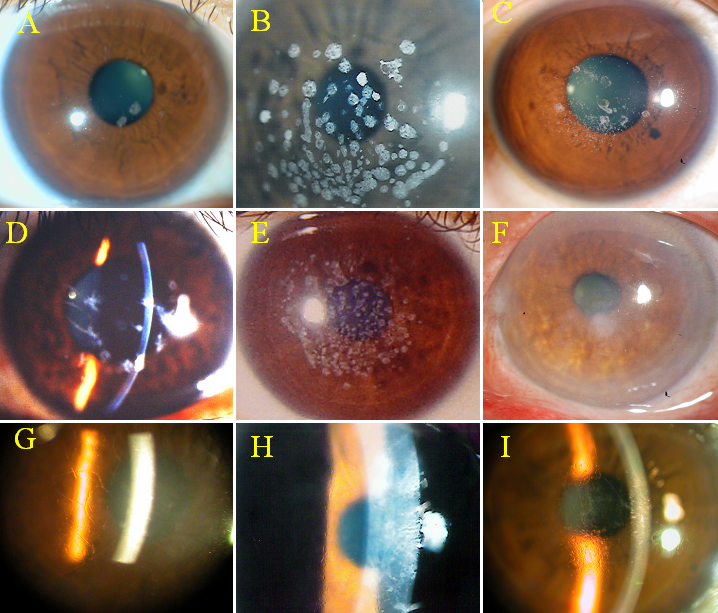
 Figure 3 of
Hou, Mol Vis 2012; 18:362-371.
Figure 3 of
Hou, Mol Vis 2012; 18:362-371.