Figure 6. Representative confocal
slices of Z-series showing Giemsa and PI staining in chicken
ossicles. The whole anterior halves of eyes from chicken embryos
were stained with both Giemsa and propidium iodide. A:
Patterns of the microporosities of chicken scleral ossicles (E16
and E20). B: Features of nuclei in chicken scleral
ossicles (E16 and E20). C: Profiles of the ossicle
microporosities containing nuclei, indicating that an individual
nucleus resides in each bone microporosity (arrows). Scale bar,
50 µm.
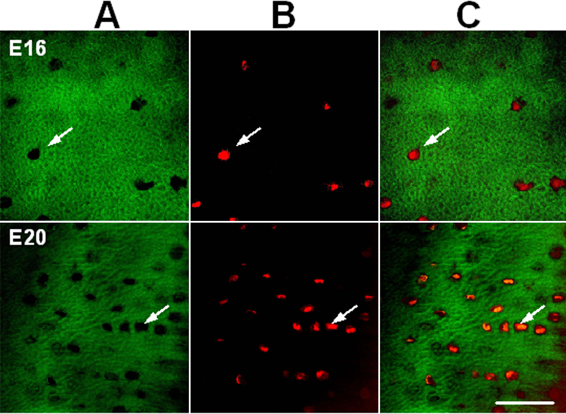
 Figure 6
of Zhang, Mol Vis 2012; 18:348-361.
Figure 6
of Zhang, Mol Vis 2012; 18:348-361.