Figure 5. FESEM images showing the
bone microporosities in the surface of chicken scleral ossicles.
A and B: Features of the bone microporosities of
E15 chicken scleral ossicles (arrow). C and D:
Patterns of the bone microporosities of E18 chicken scleral
ossicles (arrow).
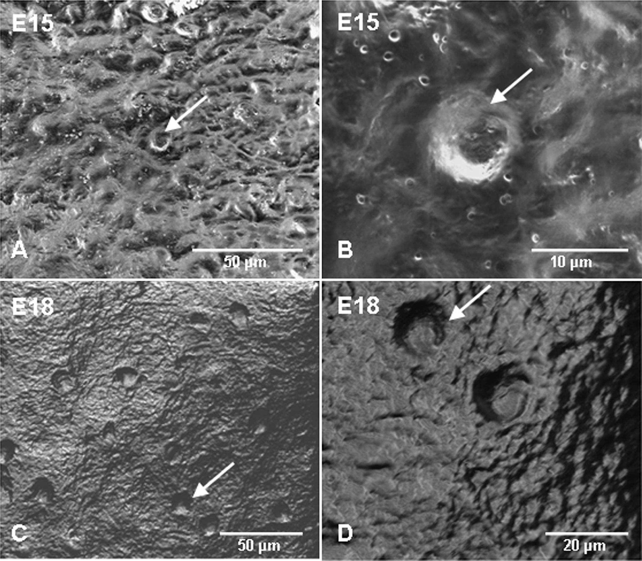
 Figure 5
of Zhang, Mol Vis 2012; 18:348-361.
Figure 5
of Zhang, Mol Vis 2012; 18:348-361.