Figure 1. The morphology of
developing chicken scleral ossicles imaged using fluorescence
stereomicroscopy. A: The development of chicken scleral
ossicles at stages E10 to E21. B: The details of
ossification, including the overlapping of adjacent ossicles
along the dorsal quadrant of the ring. A: Scale bar: 1
mm; B: Scale bar: 0.5 mm.
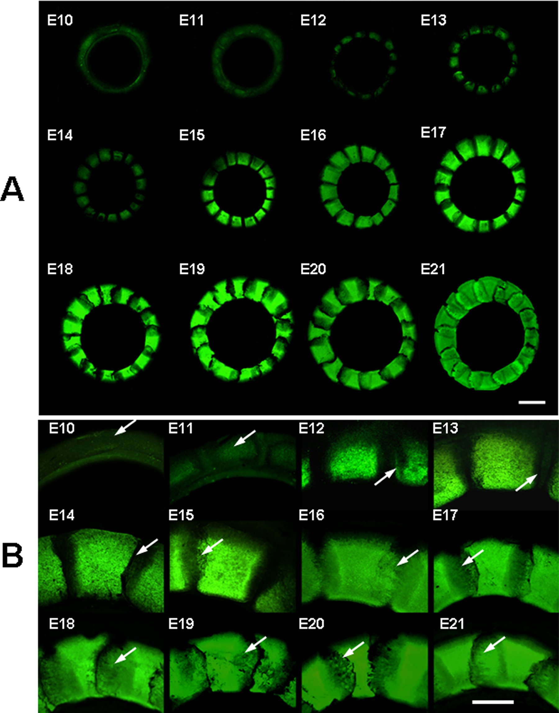
 Figure 1
of Zhang, Mol Vis 2012; 18:348-361.
Figure 1
of Zhang, Mol Vis 2012; 18:348-361.