Figure 2. K5 specifically bound to
retinal Müller cells. A: Rat Müller cells were incubated
with increasing concentrations of 125I-labeled K5
for 1 h followed with thorough washing with PBS. Then 125I-K5
bound to the cells were quantified using a γ-counter (mean±SD,
n=3). The binding of 125I-K5 to Müller cells
appeared concentration-dependent and saturated above 100 nm of
K5. B: The cells were incubated with 50 nM of 125I-
K5 and increasing concentrations of unlabeled K5 at 37 °C
for 1 h, washed with PBS, and 125I-K5 on the cells
was quantified by γ-counting (mean±SD, n=3). C: The
cells were incubated with 50 nM of 125I- K5 and
increasing concentrations of unlabeled angiostatin under the
same conditions. After thorough washing, 125I-K5 on
the cells were quantified (mean±SD, n=3). The binding of 125I-K5
to Müller cells was competed off by increasing concentrations of
unlabeled K5 but not by angiostatin.
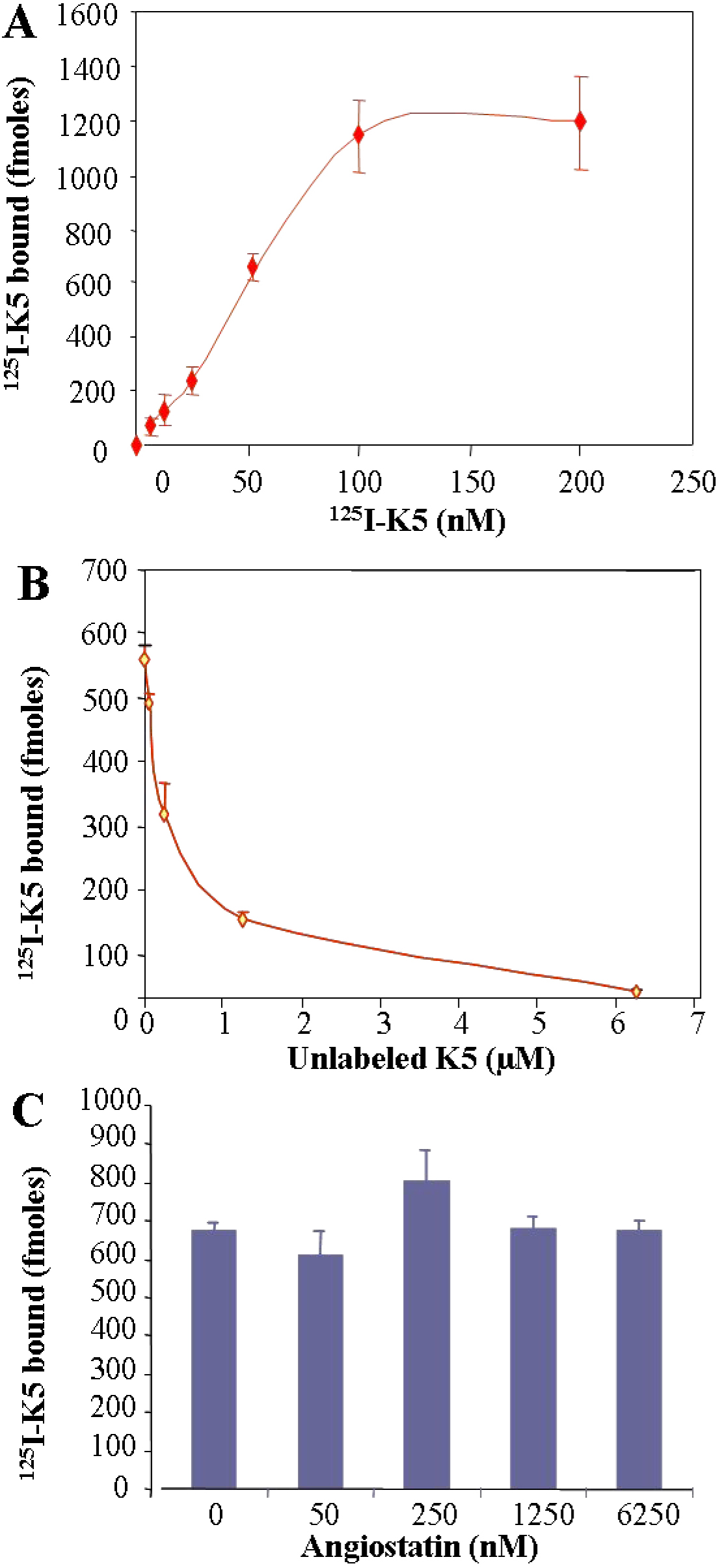
 Figure 2
of Ma, Mol Vis 2012; 18:330-336.
Figure 2
of Ma, Mol Vis 2012; 18:330-336.