Figure 3 of
Li, Mol Vis 2012; 18:317-329.
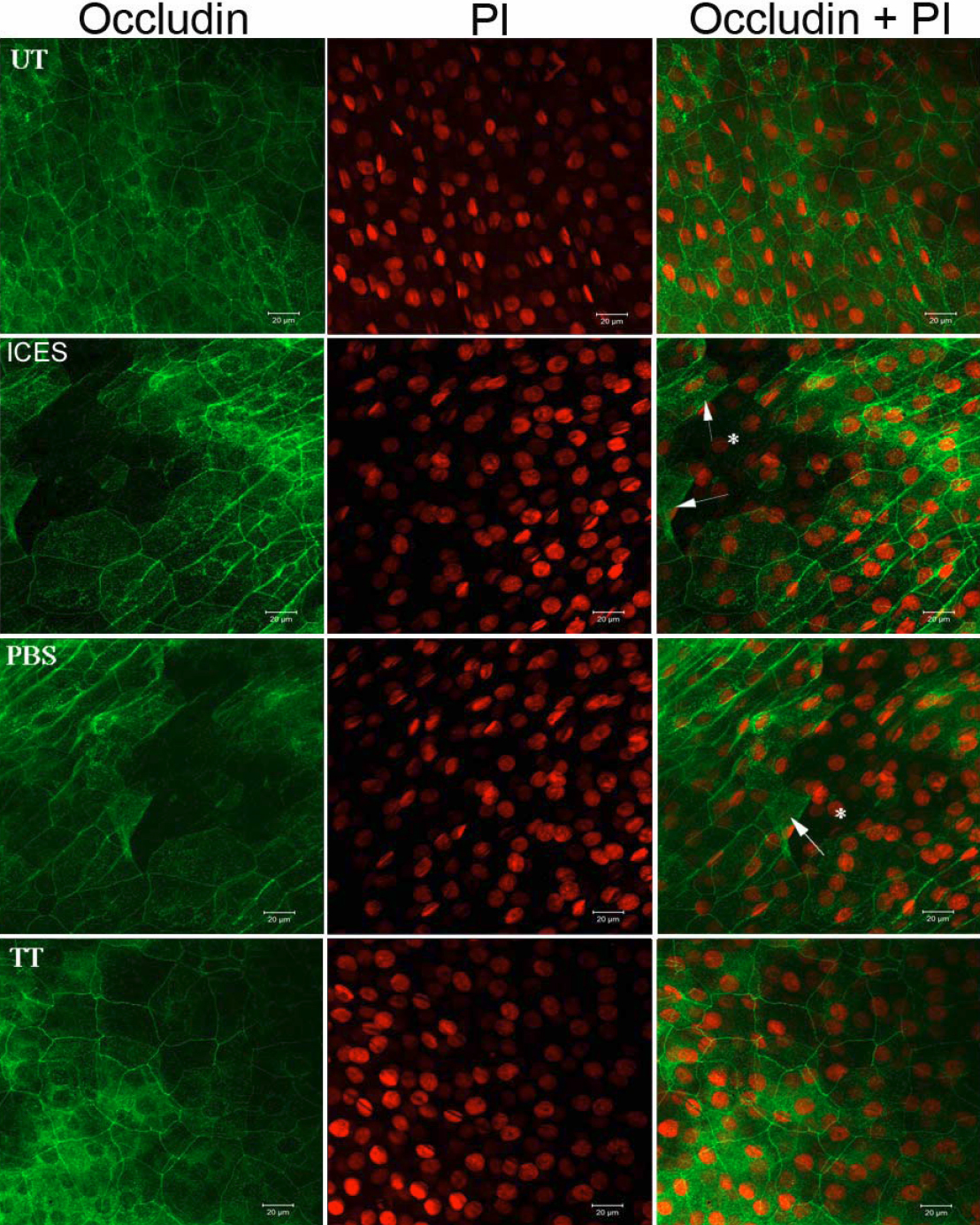
Figure 3.
Immunofluorescent staining of whole-mounted corneas stained for occludin. Arrows: desquamating apical epithelial cells; asterisk: holes resulting from the detached cells.
 Figure 3 of
Li, Mol Vis 2012; 18:317-329. Figure 3 of
Li, Mol Vis 2012; 18:317-329.
Figure 3 of
Li, Mol Vis 2012; 18:317-329. Figure 3 of
Li, Mol Vis 2012; 18:317-329.