Figure 6 of
Zhou, Mol Vis 2012; 18:309-316.
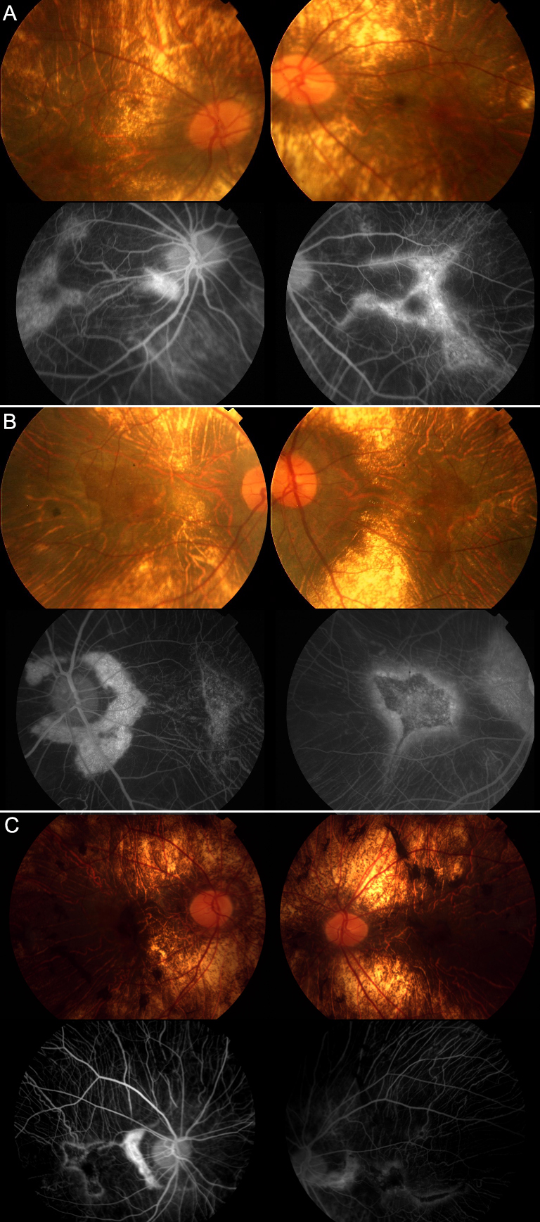
Figure 6.
Fundus photographs and fluorescein fundus angiography from choroideremia (CHM) patients. Characteristic retinal and choroidal atrophy and relatively preserved macular area are revealed. (
A
) F1-III-1, (
B
) F2-III-2, and (
C
) F3-III-2.
 Figure 6 of
Zhou, Mol Vis 2012; 18:309-316.
Figure 6 of
Zhou, Mol Vis 2012; 18:309-316.