Figure 4. Western blot results from
the family 1 show the absence of Rep-1 protein in the patient
(line 2) and a reduced amount of Rep-1 in the female carrier
(Line 1). Lane 3 is the normal brother of the patient. A:
Mouse anti-REP-1, clone 2F1 demonstrated the absence of Rep-1
protein in the patient and a reduced amount of Rep-1 protein in
the female carrier. B: β-actin antibody was used as a
loading control to ensure an adequate protein sample in each
lane.
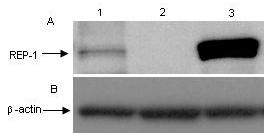
 Figure 4
of Zhou, Mol Vis 2012; 18:309-316.
Figure 4
of Zhou, Mol Vis 2012; 18:309-316.