Figure 3. Representative results of phosphate-buffered saline (PBS; A, E) blank-nanoparticles (NPs; B, F) pigment epithelium-derived factor (PEDF; C, G) and PEDF-NP (D, H) treated eyes double immunostained with antibodies to rod opsin (green) and GFAP (red) at 4 (A, B, C, D) and 8 weeks (E, F, G, H). ILM; inner limiting membrane, GCL; ganglion cell layer, IPL; inner plexiform layer, INL; inner nuclear layer, ONL; outer
nuclear layer. RPE: retinal pigment epithelium. A bar represents 50 µm.
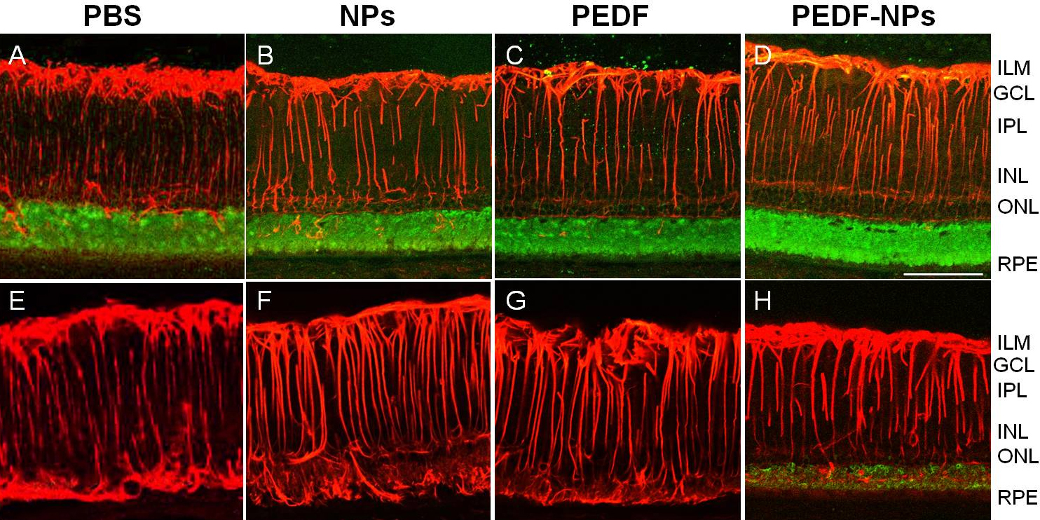
 Figure 3 of
Akiyama, Mol Vis 2012; 18:3079-3086.
Figure 3 of
Akiyama, Mol Vis 2012; 18:3079-3086.