Figure 2. Morphologic rescue of Royal College of Surgeons (RCS) rats. Representative photographs 8 weeks after injection of (A) phosphate-buffered saline (PBS), (B) blank-nanoparticles (NPs), (C) pigment epithelium-derived factor (PEDF), and (D) PEDF-NPs. ILM; inner limiting membrane, GCL; ganglion cell layer, IPL; inner plexiform layer, INL; inner nuclear layer,
ONL; outer nuclear layer. RPE: retinal pigment epithelium. A bar represents 50 µm. E: Mean number of photoreceptors per 50 µm of RCS rat retina at 4 and 8 weeks after injection. Data are shown as the mean±SD
(n=4 in each group).
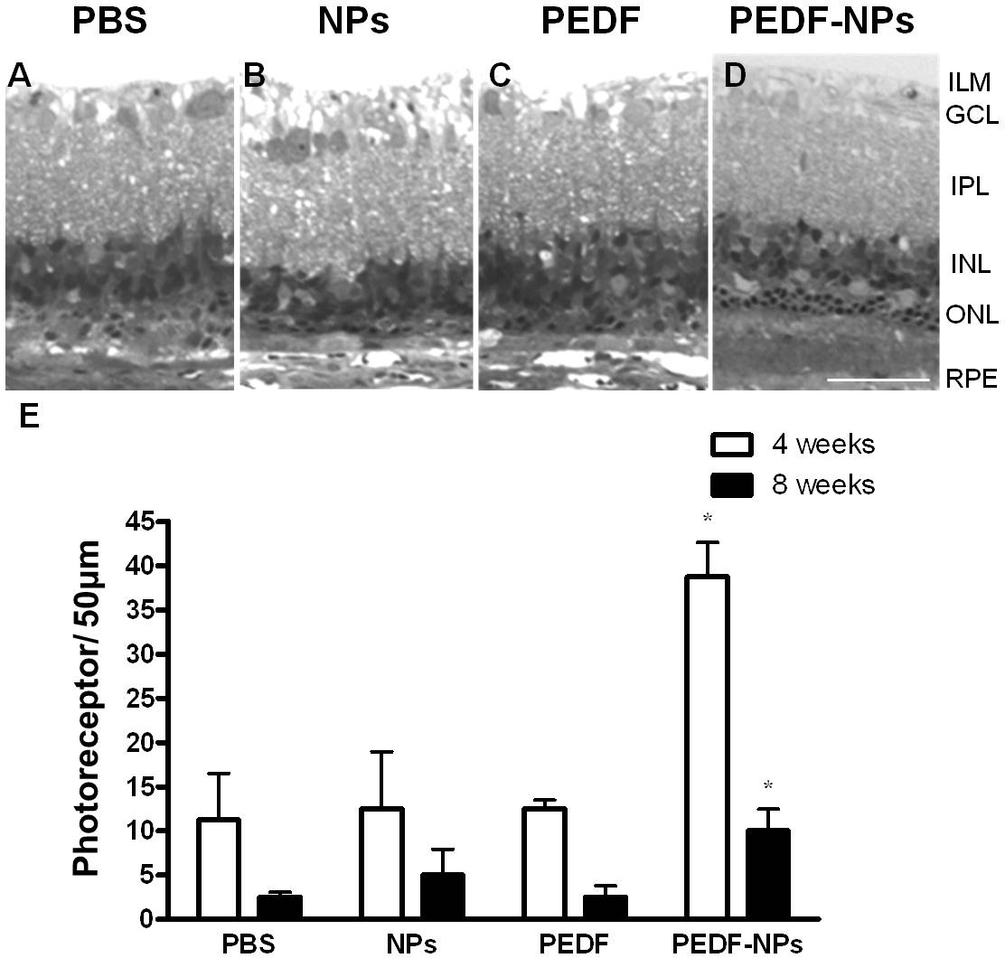
 Figure 2 of
Akiyama, Mol Vis 2012; 18:3079-3086.
Figure 2 of
Akiyama, Mol Vis 2012; 18:3079-3086.