Figure 2. Results of 1.2% agarose gel electrophoresis for amplified CFH products. Each lane represents an individual DNA sample. The number assigned to each lane corresponded to the arbitrary labels
given to each donor. “R” represents retinal DNA while “I” represents iris DNA. Thus, 1r and 1i represent retinal and iris
DNA, respectively, obtained from donor eye number 1. For each sample, 4 μl of PCR product (out of 25 μl) was used and ran
at 70 V for 30 min in 1% agarose gel. A: Every retinal DNA sample yielded detectable PCR product. B: The PCR products from the iris DNA samples were less prominently detected than retinal DNA. Some samples did not yield detectable
products. Retinal DNA bands are significantly brighter than the iris DNA bands, suggesting greater yields. Note: M=Marker
(1 kb DNA ladder, Invitrogen); NTC=non-template control (negative control).
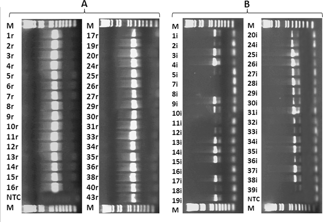
 Figure 2 of
Wang, Mol Vis 2012; 18:3049-3056.
Figure 2 of
Wang, Mol Vis 2012; 18:3049-3056.