Figure 1. Altered structure of rod spherules and cone pedicles in adult mice following postnatal-only lead exposure. Postnatal developmental
lead exposure alters the structure of rod spherules and cone pedicles in adult mice. A: A longitudinal section of a control retina showing several rod spherules and a cone pedicle. B: Longitudinal section showing two rod spherules and a cone pedicle from a lead-exposed mouse. A rod nucleus (RN) in the most
proximal row of the outer nuclear layer is not retracted. The rod spherule of the left contains a moderately swollen mitochondrion
(m) and a swollen cisterna of agranular endoplasmic reticulum (white arrow). One of the lateral horizontal cell processes
(h) of the invaginating synapse is quite swollen. The rod spherule of the right contains a mildly swollen mitochondrion (m),
and one lateral horizontal cell process (h) is quite swollen. A larger more electron lucent cone pedicle contains a moderately
swollen mitochondrion. The invaginating processes of the lateral horizontal cells and a central bipolar cell also are swollen.
Scale bar=1 micron. C: A longitudinal section from a different lead-exposed mouse showing two rod spherules and a cone pedicle. In the spherules
and pedicles, the mitochondria and cisterna of the agranular endoplasmic reticulum (white arrow) adjacent to the mitochondrion
(m) are often swollen. Similarly, the lateral horizontal cell processes (h) of the invaginating synapse can be swollen. A
synaptic ribbon (black arrow) is visible. The larger more electron lucent cone pedicle appears to have swollen invaginating
synapses. Gap junctions between the large cone pedicle and the adjacent rod spherule remain (solid black arrowheads). Scale
bar=1 micron. D: A longitudinal section from a Bcl-xL/lead mouse showing a rod spherule and a cone pedicle. Bcl-xL overexpression blocked
most of the lead-induced mitochondrial and neuronal dendritic process swelling. Scale bar=1 micron.
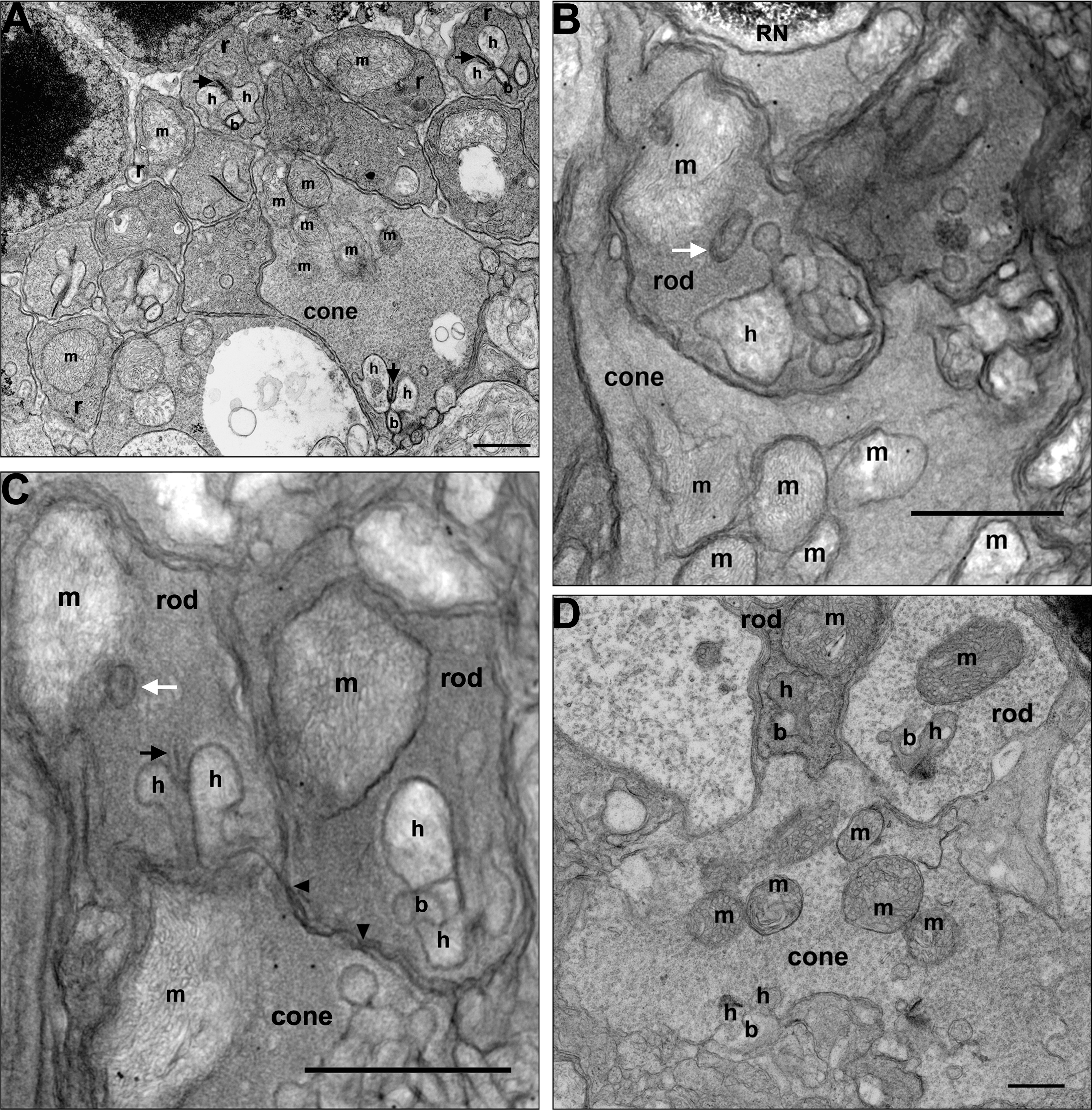
 Figure 1 of
Perkins, Mol Vis 2012; 18:3029-3048.
Figure 1 of
Perkins, Mol Vis 2012; 18:3029-3048.