Figure 5. Fundus photograph and optical coherence tomography image of the right eye for II:1 (ADRP-LG). Fundus examination showed typical
high myopic fundus changes including tilting of the optic disc, myopic conus, and tessellated fundus. Optical coherence tomography
image revealed relatively normal macular lamination.
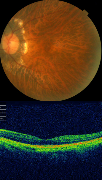
 Figure 5 of
Xu, Mol Vis 2012; 18:3021-3028.
Figure 5 of
Xu, Mol Vis 2012; 18:3021-3028.