Figure 4. Fundus photographs (A and F) and optical coherence tomography images (C, D, H, and I) of both eyes for III:2 (ADRP-XL). Panel B and G indicated red-free fundus images. Panel E and J indicated the calculating results of macular thickness and total volume in the given areas. Right eye: panel A-E. Left eye: panel F-J. No signs of RP can be seen.
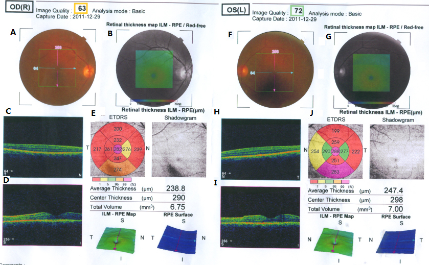
 Figure 4 of
Xu, Mol Vis 2012; 18:3021-3028.
Figure 4 of
Xu, Mol Vis 2012; 18:3021-3028.