Figure 3. Fundus photographs(top panel) and optical coherence tomography images (lower panel) of four probands with mutations in the
PRPF31 gene: A and B: III:1 (ADRP-HT); C and D: III:9 (ADRP-LG); E and F: III:1 (ADRP-LLN); G and H: IV:1 (ADRP-XL). Typical retinitis pigmentosa appearance of the fundus can be seen (A, C, E, and G). Optical coherence tomography images (B, D, F, and H) reveal relatively preserved foveal lamination.
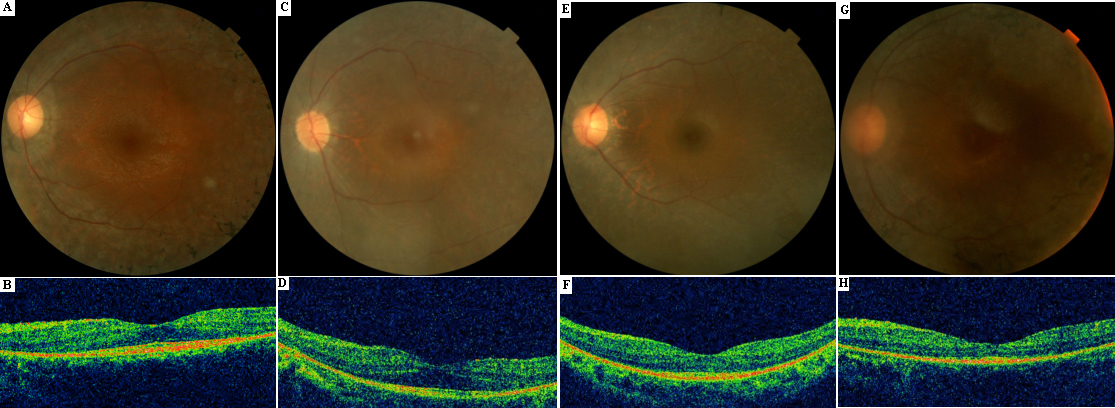
 Figure 3 of
Xu, Mol Vis 2012; 18:3021-3028.
Figure 3 of
Xu, Mol Vis 2012; 18:3021-3028.