Figure 2. Fundus appearance of two patients with retinitis pigmentosa. A: Fundus appearance of the right eye of patient III:6 in family 83 shows mild atrophic retinal pigment epithelial changes
and a few pigments in the inferior periphery fundus. B: Fundus appearance of the right eye of patient IV:5 in family 112 presents atrophic retinal pigment epithelial changes, attenuation
of the retinal vessels, and irregular pigment clumps in the retina. Numbers in the upper-left corners are the patients’ birth
dates.
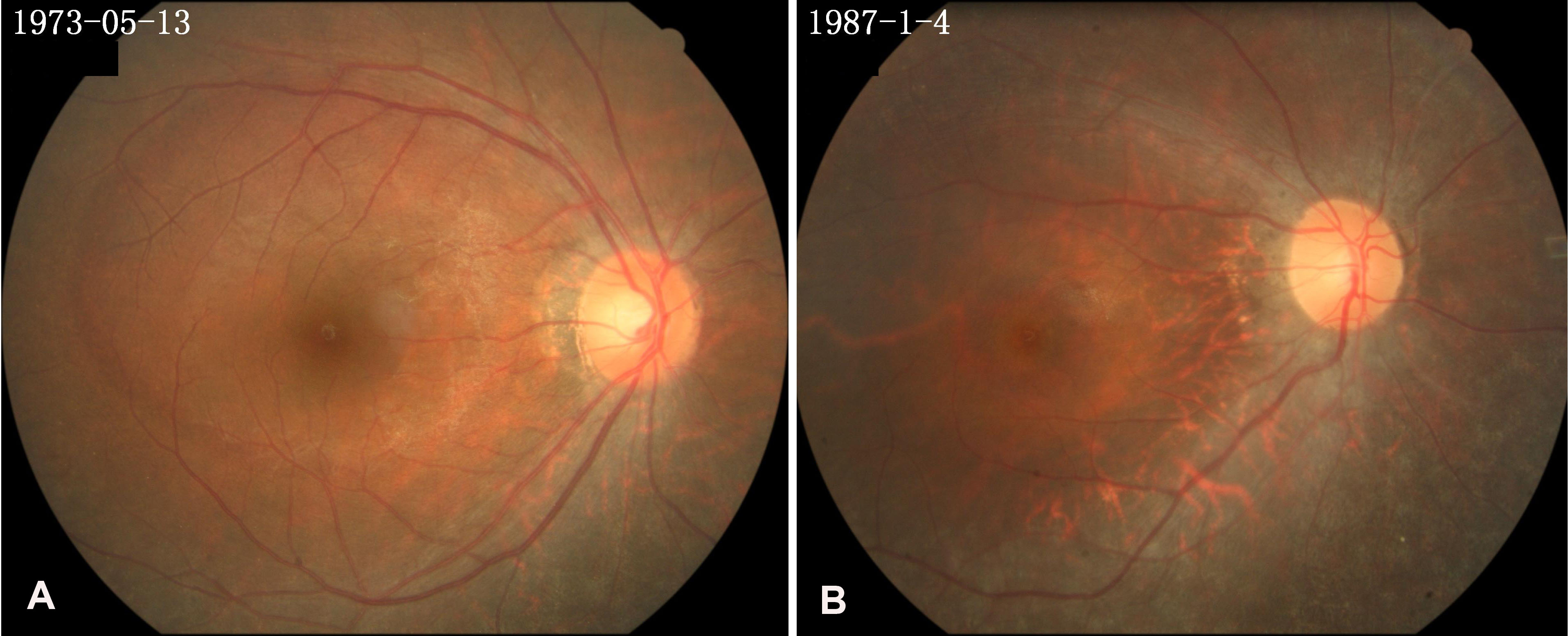
 Figure 2 of
Pan, Mol Vis 2012; 18:3013-3020.
Figure 2 of
Pan, Mol Vis 2012; 18:3013-3020.