Figure 2. OCT and ERG results of patient 2. A: OCT image of the right eye showing foveal cystic retinoschisis. B: OCT image of the left eye showing foveal atrophy. C, D: Standard scotopic response of full-field ERG of the right and left eye showing decreased b-wave amplitude
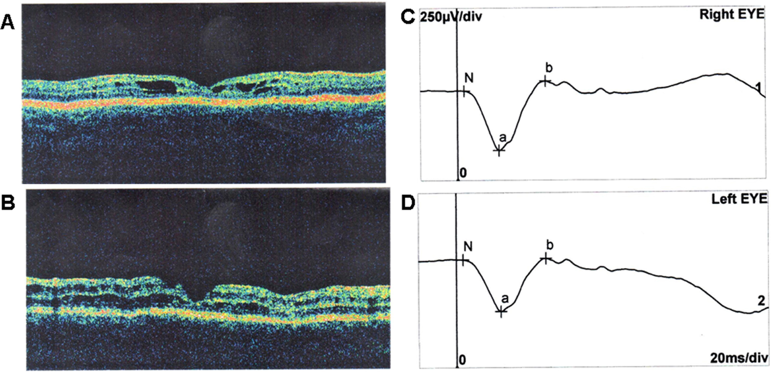
 Figure 2 of
Skorczyk, Mol Vis 2012; 18:3004-3012.
Figure 2 of
Skorczyk, Mol Vis 2012; 18:3004-3012.