Figure 7. Overexpression of proNGF caused retinal microvascular degeneration. A: Western blot (WB) analysis of retinal lysate showed significant increase in proNGF (32 kDa) expression in rats electroporated
with proNGF123 as compared with those electroporated with pGFP (n=4). The asterisk represents significant difference as compared
with the control group at p<0.05. B: Representative images for retinal trypsin digests stained with periodic acid-Schiff and hematoxylin (PASH) showing acellular
capillaries (arrows). C: Statistical analysis of the number of acellular capillaries showed no significant difference after 1 week of proNGF overexpression
(n=4). Analyses after 4 weeks showed significant increases in the number of acellular capillaries in retinas injected with
pGFP-proNGF123 when compared to the pGFP control group (n=4; * p value <0.05).
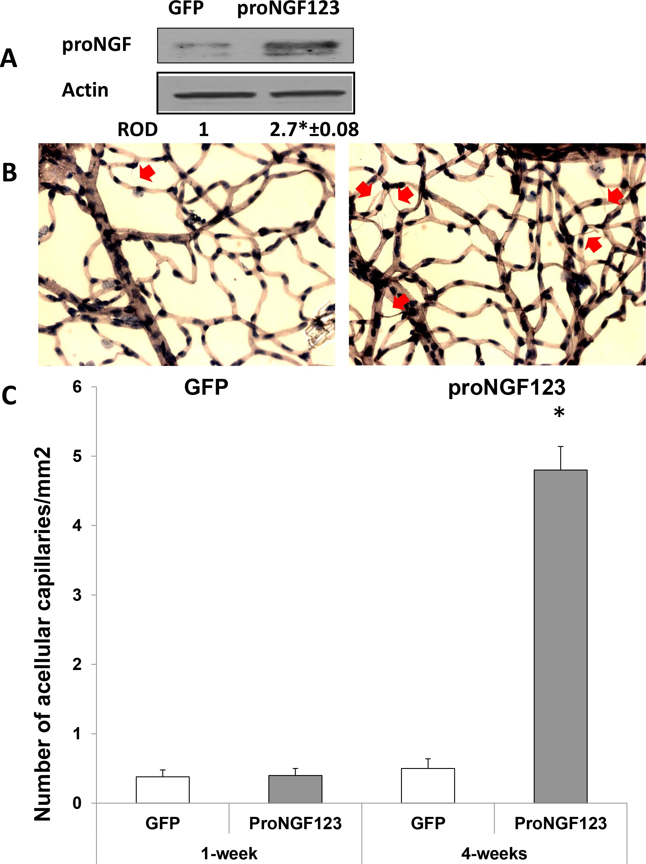
 Figure 7 of
Matragoon, Mol Vis 2012; 18:2993-3003.
Figure 7 of
Matragoon, Mol Vis 2012; 18:2993-3003.