Figure 5. Overexpression of proNGF reduced the number of cells in the ganglion cell layer. A, B: Representative images and statistical analysis of rat retina sections stained with hematoxylin and eosin showing a reduction
in number of neuronal cells in the ganglion cell layer in rats injected with p-GFP-proNGF123 as compared with pGFP controls
in the central and posterior retina (n=4, 200X magnification). C, D: Representative images and statistical analysis of the number of RGC normalized to the retinal length. RGCs were counted
as Brn3a-positive cells (Red) and DAPI (Blue). The results showed a significant reduction in RGC number in rats injected with
p-GFP-proNGF123 as compared with pGFP controls. The asterisk represents significant difference as compared with the control
group at p<0.05. IPL, inner plexiform layer; INL, inner nuclear layer; ONL, outer nuclear layer.
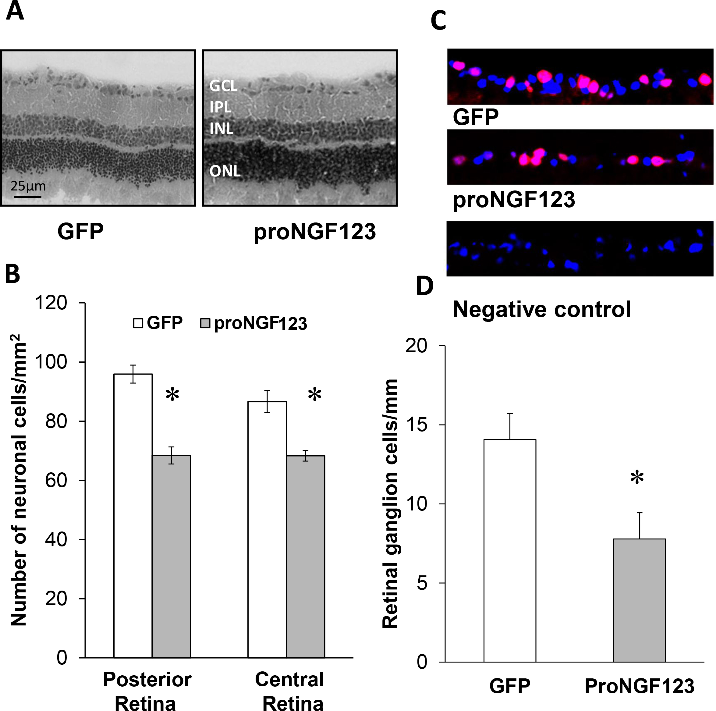
 Figure 5 of
Matragoon, Mol Vis 2012; 18:2993-3003.
Figure 5 of
Matragoon, Mol Vis 2012; 18:2993-3003.