Figure 4. Overexpression of proNGF caused neuronal cell death A: Representative images of retina sections show TUNEL-fluorescein-positive cells mainly within the ganglion cell layer (GCL)
and inner nuclear layer (INL) of rats injected with p-GFP-proNGF123 as compared with the pGFP control (200X magnification).
B: Quantitative assessment of total number of TUNEL–horseradish peroxidase (HRP)-positive cells in the flatmounted retina showing
5.5-fold increase of cell death in retinas from rats injected with proNGF compared to pGFP controls (n=5–6). * represents
significant difference as compared with the control group at p<0.05. IPL, inner plexiform layer; ONL, outer nuclear layer.
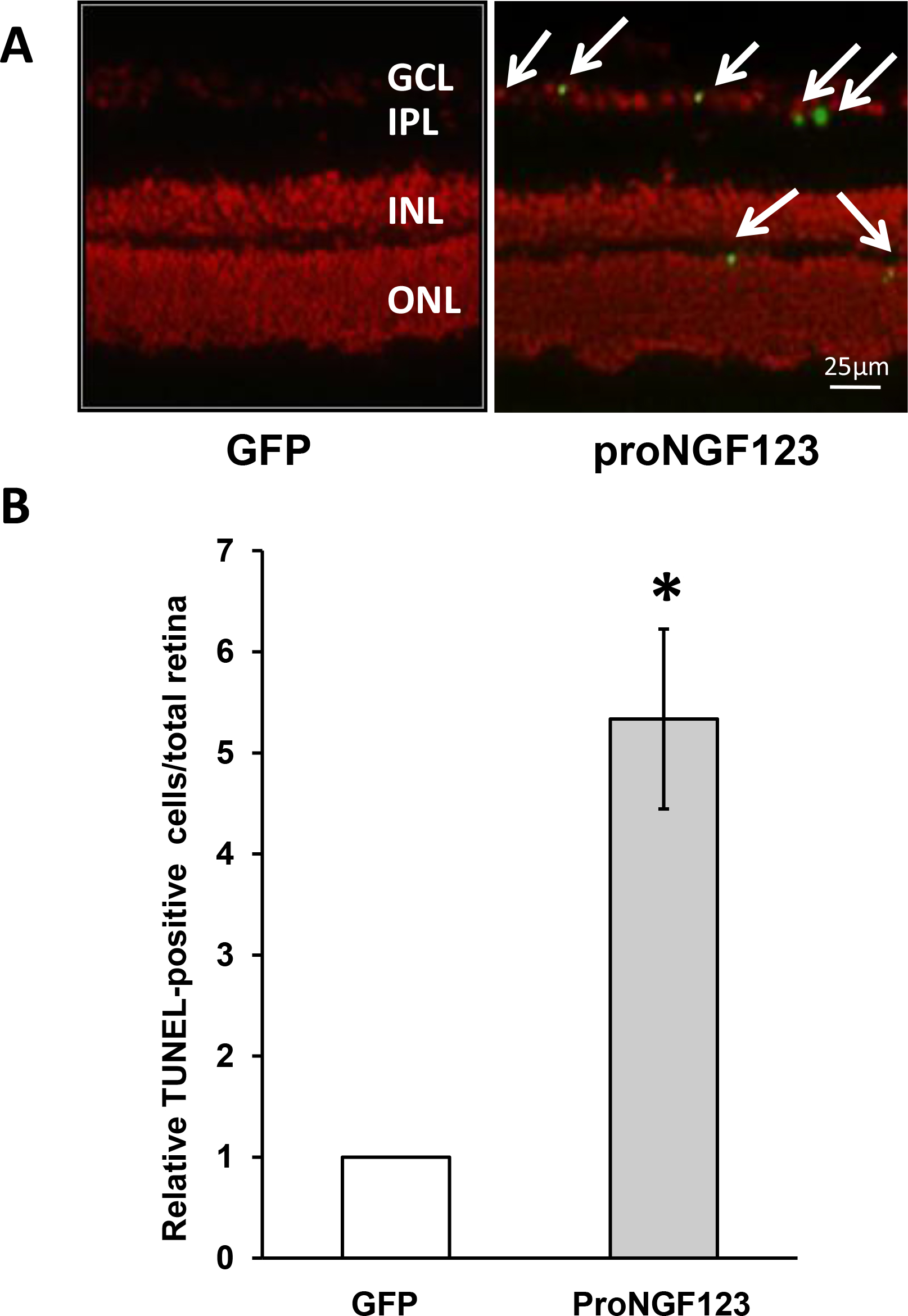
 Figure 4 of
Matragoon, Mol Vis 2012; 18:2993-3003.
Figure 4 of
Matragoon, Mol Vis 2012; 18:2993-3003.