Figure 3. Microglia transduced with CC-chemokine receptor 2 (CCR2)-green fluorescent protein (GFP) lentivirus express microglia marker.
Some cells are spindle shaped and a few are ramified; both shapes represent the “silent” state of microglia, while approximately
50% of primary microglia showed amoeboid or round shapes, indicating a state of activation. This culture composition is very
similar to that of the untreated primary microglia. A: Microglia transduced with CCR2-GFP are shown under light microscopy. B: Microglia transduced with CCR2-GFP express green fluorescence. C- H: Microglia transduced with CCR2-GFP express microglia marker ED1 and Iba1 in vitro.
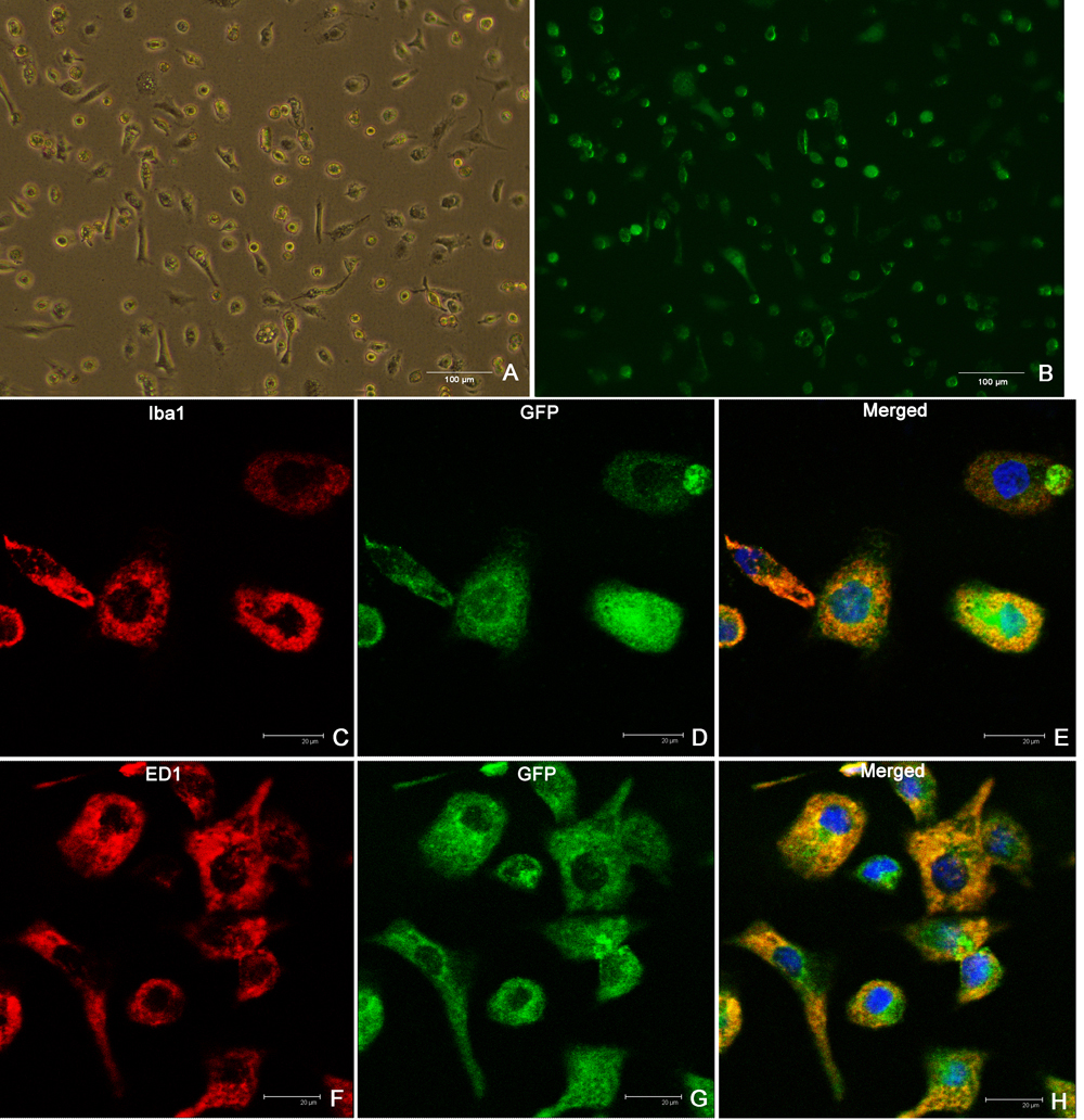
 Figure 3 of
Jiang, Mol Vis 2012; 18:2982-2992.
Figure 3 of
Jiang, Mol Vis 2012; 18:2982-2992.