Figure 2. Identification of microglia transduced with green fluorescent protein (GFP)-tagged CCR2 (CCR2-GFP) lentivirus. A: Flow cytometric analysis showed untreated primary microglia were used as controls. B: Transduction efficiency was analyzed by flow cytometry for GFP fluorescence and reached 97.6%. C: Western blotting shows that microglia transduced with CCR2-GFP (MGC-ccr2) expressed more CCR2 than microglia transduced
with GFP (MGC-gfp). TTF was tail tip fibroblast of mouse and was a negative control. D-F: Microglia transduced with CCR2-GFP express CCR2 and GFP.
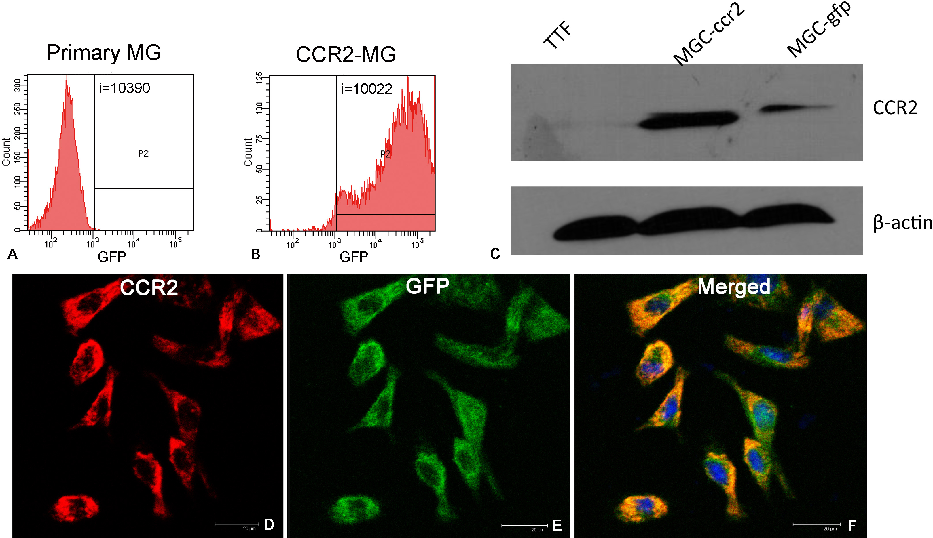
 Figure 2 of
Jiang, Mol Vis 2012; 18:2982-2992.
Figure 2 of
Jiang, Mol Vis 2012; 18:2982-2992.