Figure 4. Enhanced human retinal endothelial cell apoptosis after ARF1 inhibitor treatment. A: Representative reverse transcription polymerase chain reaction (RT–PCR) of ARF1 expression on cultured human retinal endothelial
cells (HRECs) is shown here. B: HRECs were cultured in six-well plates in Dulbecco’s modified Eagle’s medium containing 10% fetal calf serum (FCS) alone
or ARF1 inhibitor (14 μM) for 24 h. The cell apoptosis rates in each sample were determined as described in the Methods section.
The results from five independent experiments are shown. Each symbol represents the percent of cell apoptosis in each group.
The Student t test (two-tailed) was used.
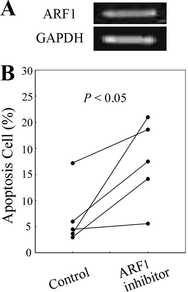
 Figure 4 of
Dai, Mol Vis 2012; 18:2947-2953.
Figure 4 of
Dai, Mol Vis 2012; 18:2947-2953.